
Transform raw information from OCT scans into structured, research-ready data.
- Work with OCT scans from 9 Major OCT Manufacturers
- Enable historical data analysis of 40+ retinal biomarkers relevant to reserach of 30+ retinal conditions
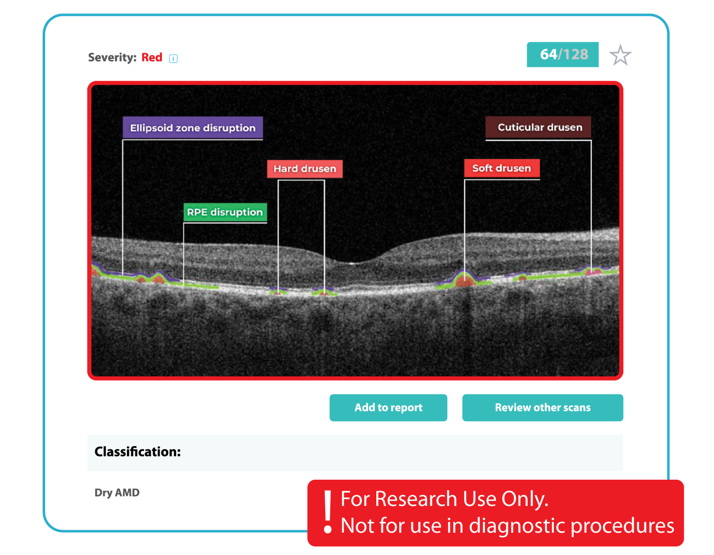
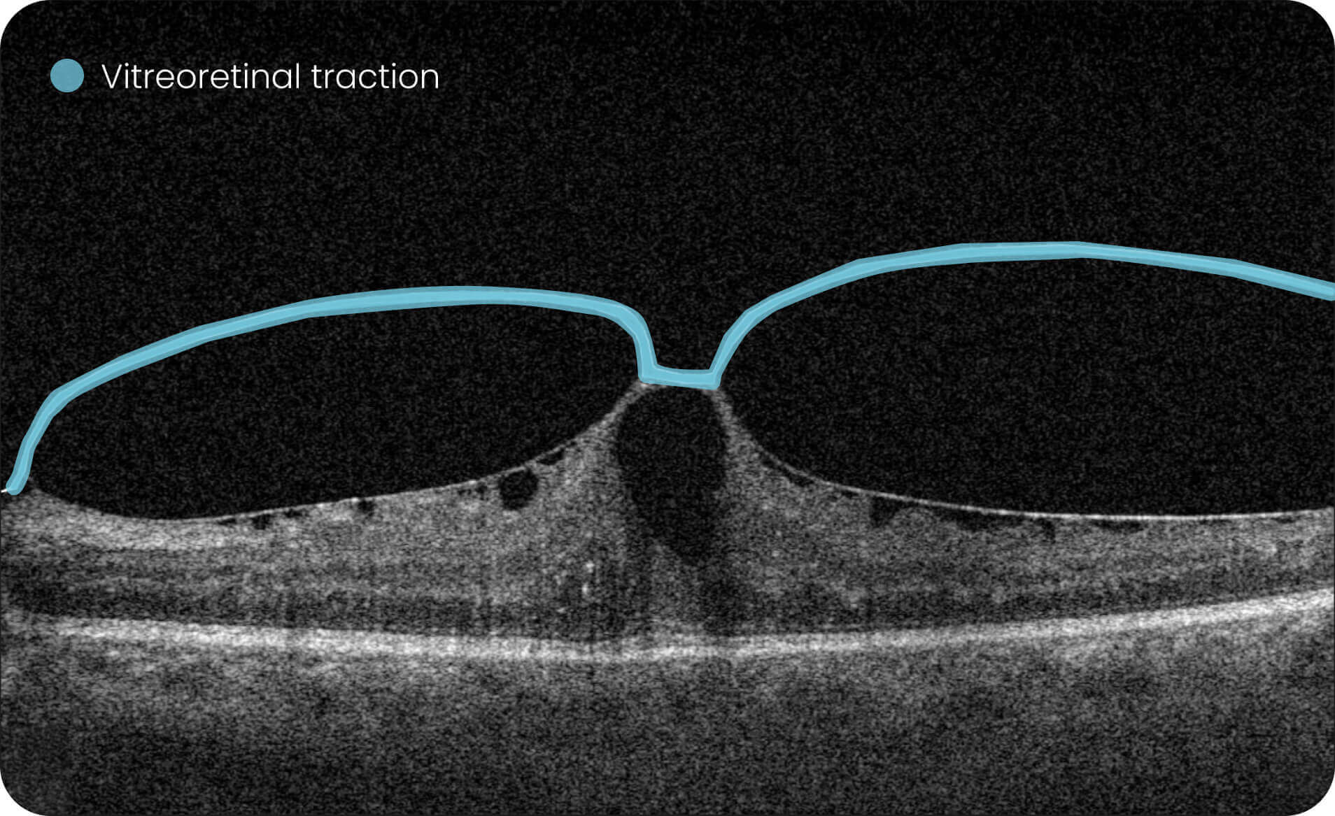
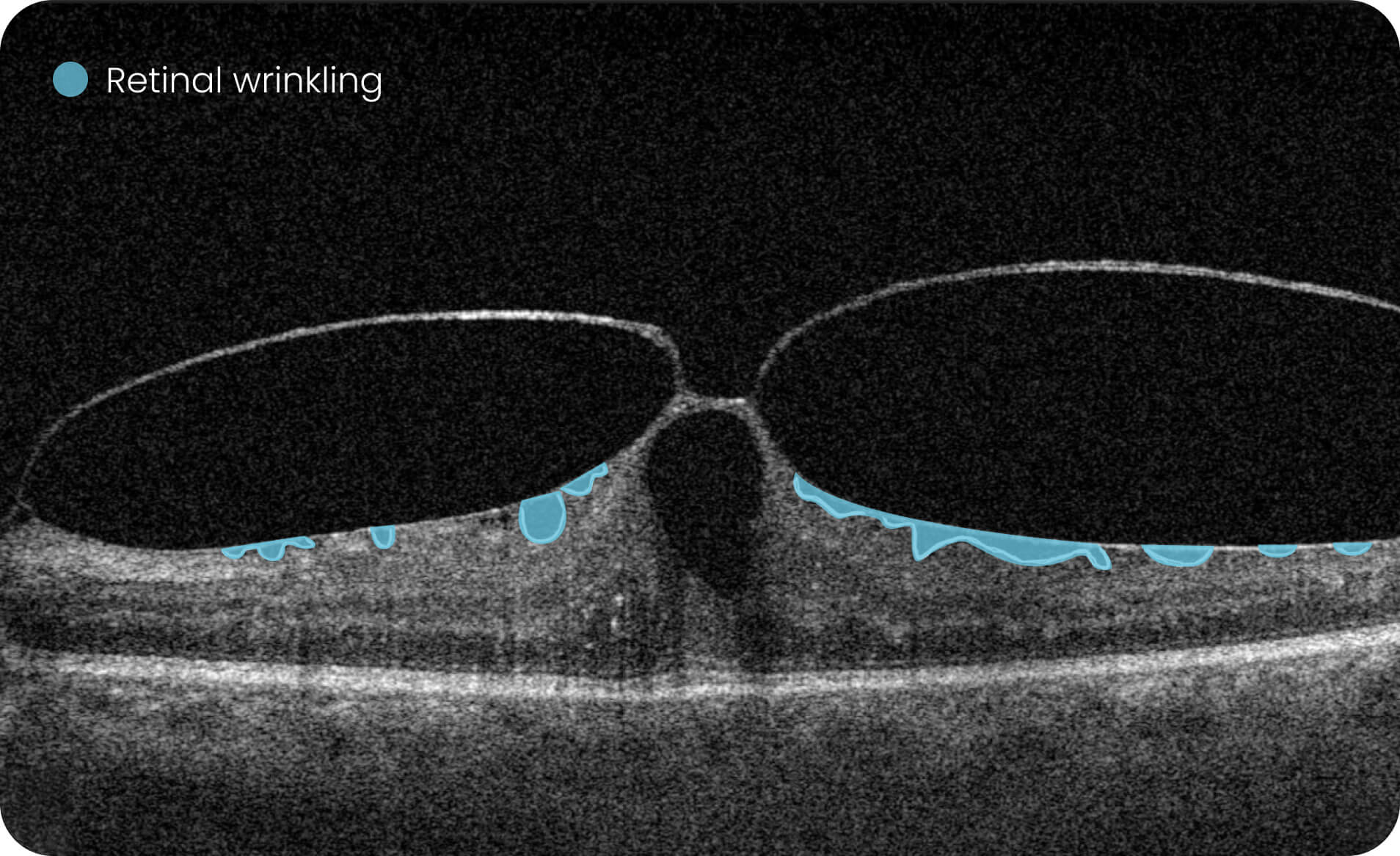
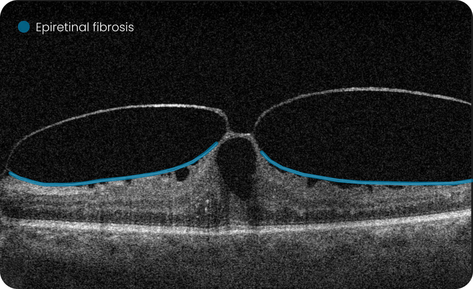
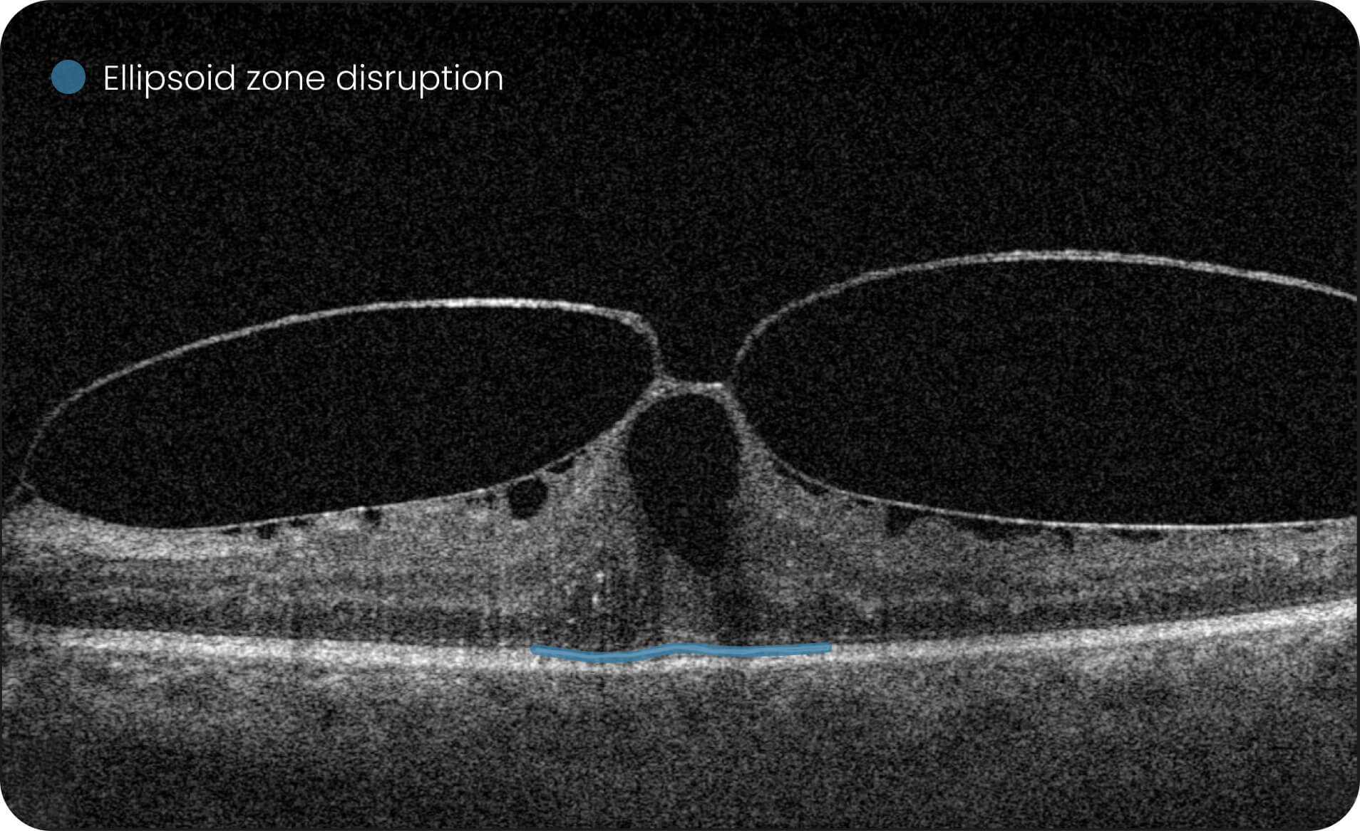
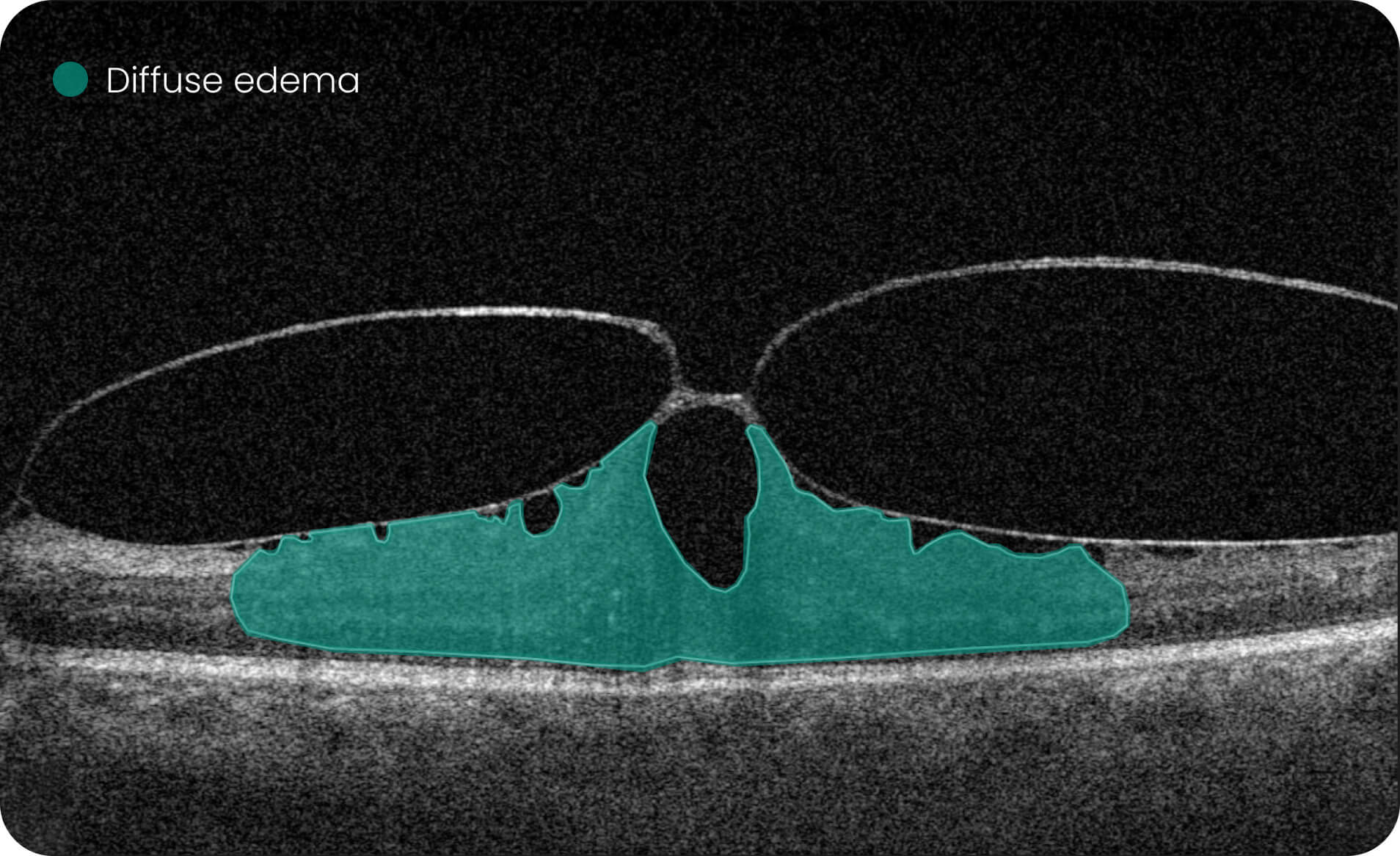
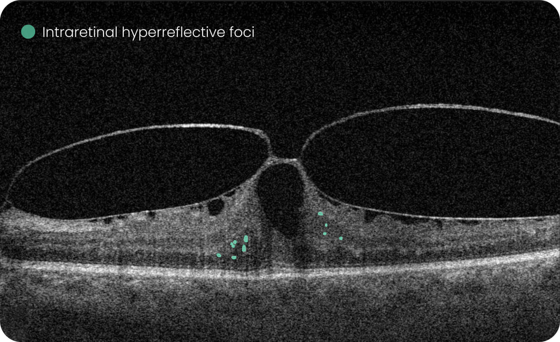
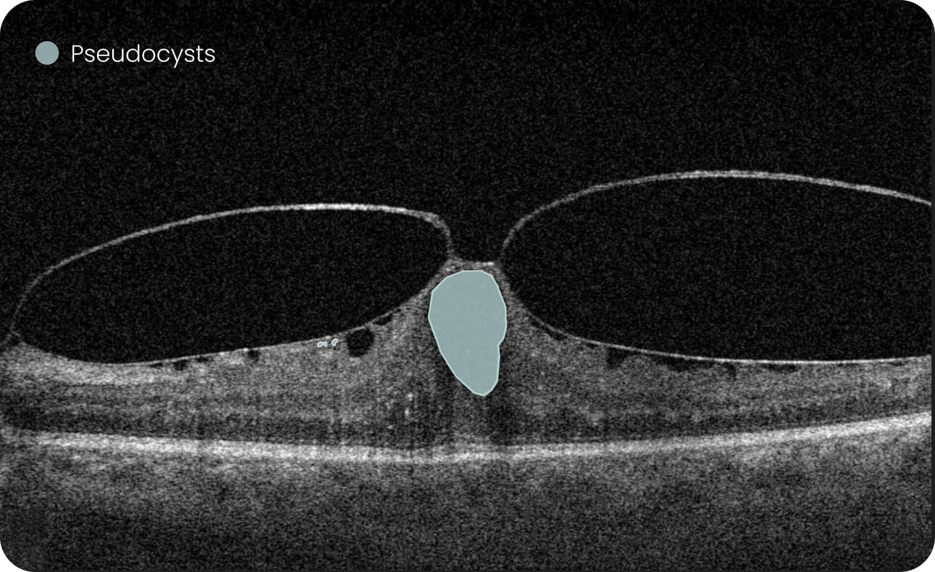
- Quantify and analyze 40+ retinal biomarkers relevant to reserach of 30+ retinal conditions
For Research Use Only.Not for use in diagnostic procedures. - Fuel ophthalmic research with 40+ retinal biomarkers relevant to reserach of 30+ retinal conditions
For Research Use Only.Not for use in diagnostic procedures. - Study progression patterns of 40+ retinal biomarkers relevant to reserach of 30+ retinal conditions
For Research Use Only.Not for use in diagnostic procedures.
Created to elevate Pharma, Optometry & Ophthalmology.


Short video about Altris IMS
End-to-End Encryption
The security of patients’ data is our top priority:
we are GDPR compliant, all data is encrypted, CE-certified, and FDA-cleared (510k) as and IMS system.
 Helen Cooper
Project Manager
Helen Cooper
Project Manager

 Dr.Samuel Minaker
Director of Clinical Research
Dr.Samuel Minaker
Director of Clinical Research

 Alisdair Buchanan
Optometry Owner, UK
Alisdair Buchanan
Optometry Owner, UK
- Explore 40+ retinal biomarkers studied across major retinal research areas.
- Work with ROU AI Models for Dry AMD, Wet AMD, GA, DR, DME, and CME. All the 40+ biomarkers and 30+ retina pathologies can be used for Research Use Only. Not for use in diagnostic procedures.
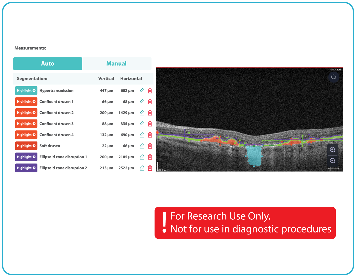
- Add manual annotations to highlight areas of reserach
- Enable vendor-neutral OCT data management. We work with Canon, Heidelberg Engineering, Nidek, Optos, Optopol, Topcon, Visionix, and Zeiss
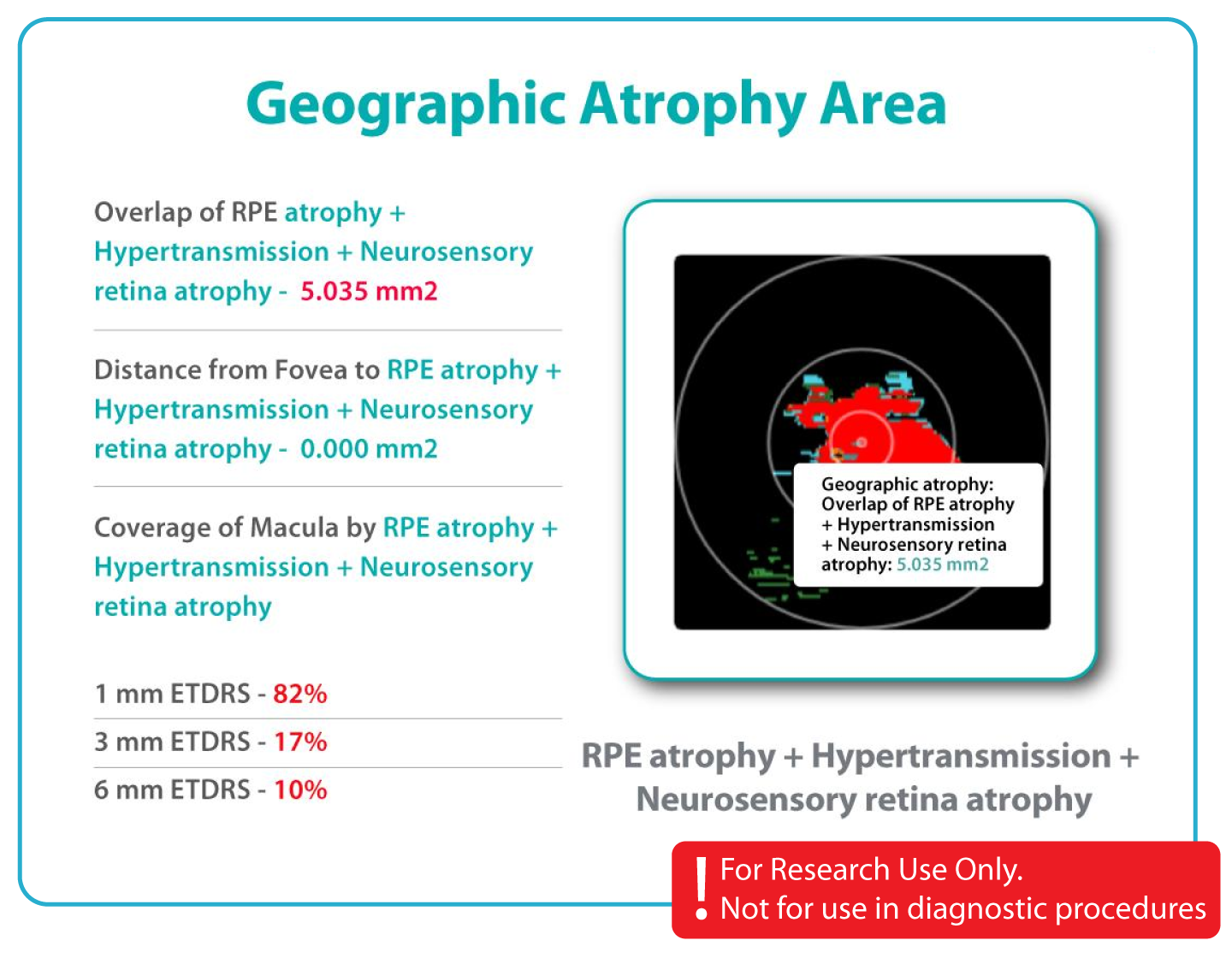
- Analyze historical data and generate clinical insights
- Store all your OCT data securely in the cloud or on-premises
-
Streamline collaboration and data sharing. Enable multiple researchers to access and annotate OCT datasets simultaneously
Transform raw OCT data into structured research-ready data.
What's the value of Altris IMS?
And how it works
Altris IMS is a web platform created by retina experts for all eye care specialists and clinicians.
We built a unique vendor-neutral IMS that works with OCT devices from 9 major manufacturers.
Detect over 40+ retina biomarkers & 30+ retina pathologies using Research Only Use AI models
Measure and track pathological changes across multiple examinations for more effective treatment decisions

Formats
DICOM format will help you to extract maximum information.

OCT equipment
Altris IMS is vendor-neutral. We work with 9 top OCT manufacturers.

OCT reports
We create comprehensible OCT reports for patients and doctors
For Pharma
For innovative approach in pharma
Start Free Trial- Centralized OCT Data Management
- Historical Data Analysis
- Research-Ready Ecosystem
- Vendor-neutral analysis of OCT scans ( 9 manufacturers)
- Data Security and Compliance
- Additional capabilities for research
- 40+ retinal biomarkers studied in research across 30+ retinal conditions. For Research Use Only. Not for diagnostic procedures.
- Quantitative exploration of 40+ biomarkers for Research Use Only. Not for diagnostic procedures.
- Reports
For eye care
IMS for Ophthalmology and Optometry
Start Free TrialWhat you get:
- Centralized OCT Data Management
- Vendor-Neutral OCT Compatibility ( 9 manufacturers)
- Secure and Compliant Data Environment
- Historical OCT Data Analysis
- Seamless Clinical Workflow Integration
- 40+ retinal biomarkers studied in research across 30+ retinal conditions. For Research Use Only. Not for diagnostic procedures.
- Quantitative exploration of 40+ biomarkers for Research Use Only. Not for diagnostic procedures.