

Mark Braddon
VP of Sales at Altris AI
Reading time
6 min read
DICOM Format: Benefits of Managing DICOM images
DICOM file format (Digital Imaging and Communications in Medicine) was developed by the American College of Radiology (ACR) and the National Electrical Manufacturers Association (NEMA) as a standard for exchanging medical images and related information across different healthcare systems. It serves as a universal language for medical imaging, enabling interoperability between various imaging devices and systems. DICOM ensures that medical images can be exchanged and viewed consistently regardless of the manufacturer or modality.
DICOM image format supports a broad range of medical imaging modalities, including X-ray, MRI, OCT, ultrasound, nuclear medicine, and more. It also covers related data, such as patient information, study details, image annotations, and results
As the DICOM format continues to evolve to keep up with advancements in medical imaging technology, our article aims to raise awareness among ophthalmologists and optometrists about the DICOM file format.
What is DICOM format? You can also watch a short video about DICOM and non-DICOM file formats.
What is DICOM file format?
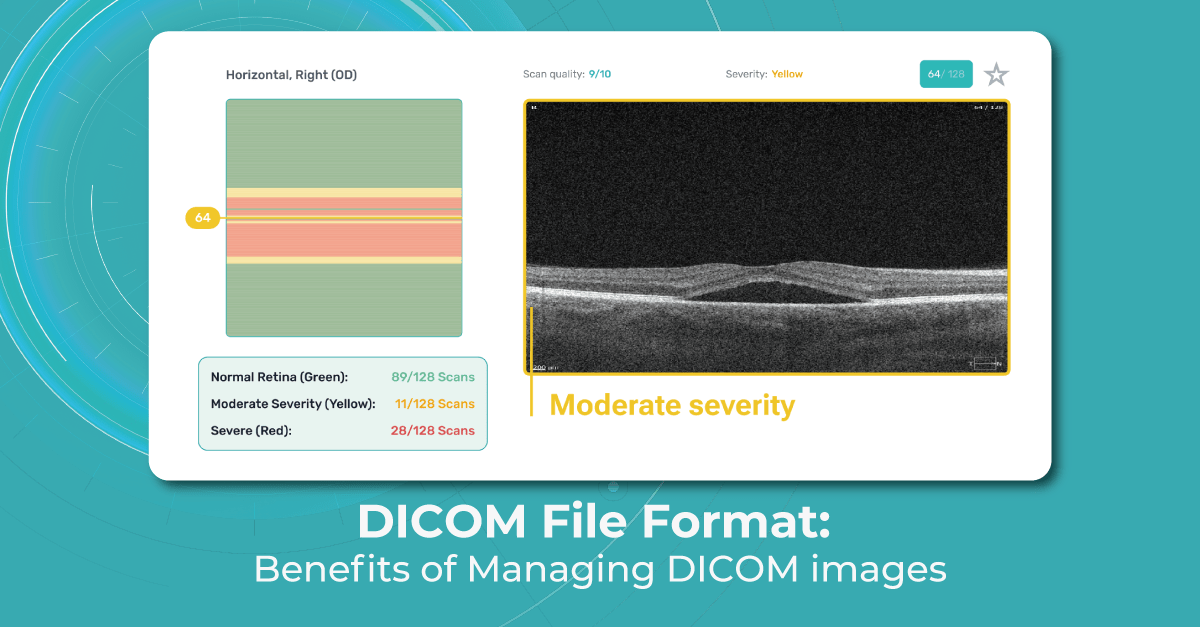
Image files that adhere to part 10 of the DICOM standard are commonly known as “DICOM format files” or simply “DICOM files,” and their file extension is “.dcm.” In ophthalmology, DICOM is a widely used file format for storing and transmitting medical images. DICOM files are used to store various types of ophthalmic images as well, including retinal images, optical coherence tomography (OCT) scans, visual field tests, and angiography images.
DICOM files consist of two main components: the header and the image data. The header contains metadata that describes the patient, study, series, and image acquisition parameters.
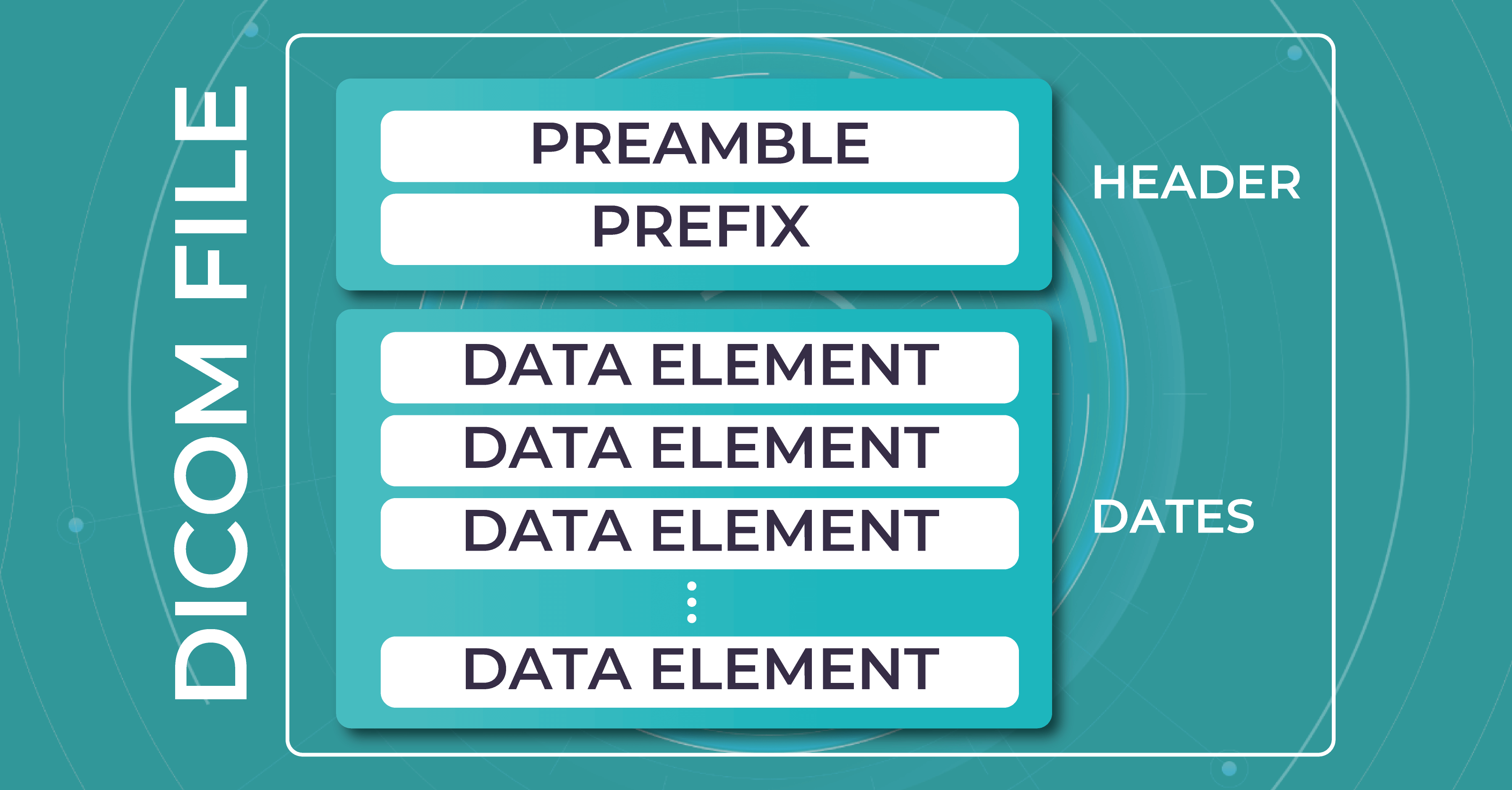
This metadata includes information such as patient demographics, image acquisition parameters (e.g., imaging modality, image orientation, pixel spacing), and any annotations or measurements made on the image. The image data itself is typically stored in a compressed format, such as JPEG or JPEG 2000, within the DICOM file.
DICOM files also support the exchange of images and associated data between different medical imaging devices and systems. This enables eye care specialists to easily share and access ophthalmic images across different platforms, such as picture archiving and communication systems (PACS), ophthalmic imaging devices, and electronic health record (EHR) systems.
By using DICOM, ophthalmologists and optometrists can efficiently store, retrieve, and analyze ophthalmic images, ensuring accurate diagnoses and effective patient care. In the next paragraphs, we will tell you more about the benefits of the DICOM file format for eye care specialists.
Benefits of DICOM format
The DICOM standard ensures interoperability between different vendors’ OCT devices and facilitates seamless data sharing and analysis. The main difference between DICOM and other image formats is that it groups information into data sets. A DICOM file consists of several tags, all packed into a single file. It stores such info as:
- demographic details about the patient
- imaging study’s acquisition parameters
- image dimensions
- matrix size
- color space
- an array of additional non-intensity information necessary for accurate image display by computers.
If you have to enter the patient’s information manually, there’s always a chance you can misspell the name or other information. However, when using a DICOM file to store patients’ information and monitor patients’ health, eye care specialists can be sure the chance of human bias is much lower.
When you work in an optometry practice or a clinic, you may spend a lot of time filling in the details every time you upload a file. And if your clinic is busy and you do 30-50 uploads daily, it could take hours. Using DICOM image format significantly speeds up the process and reduces errors.
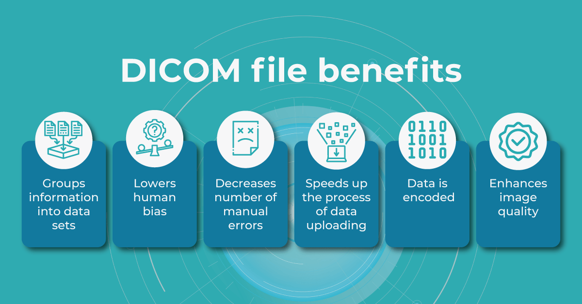
Another benefit of the DICOM image format is that the header data information is encoded within the file so that it cannot be accidentally separated from the image data.
DICOM files can be stored in a DICOM server or transmitted between DICOM-compliant systems using the DICOM network protocol (DICOM C-STORE or DICOMweb). DICOM SR (structure reporting) allows for the structured representation of measurement data and annotations in OCT images. It enables the storage of quantitative measurements, such as retinal thickness or optic nerve parameters, as structured data within the DICOM file.
In addition, eye care specialists are able to manipulate the brightness of the image when using the DICOM viewing software. Some areas of an image can be increased or decreased for a better viewing and diagnostic experience.
Is DICOM file format popular among OCT providers?
When it comes to optical coherence tomography, many OCT device manufacturers and software providers support the DICOM standard for storing and exchanging OCT images. Some of the prominent OCT providers that offer DICOM support include:
- Heidelberg Engineering is a well-known provider of OCT devices and software solutions for ophthalmology. They offer OCT devices like the Spectralis OCT, which supports DICOM connectivity. The DICOM capabilities of their systems enable seamless integration with PACS and other healthcare systems.
- Carl Zeiss Meditec is a leading manufacturer of ophthalmic devices, including OCT systems. Their OCT devices, such as the Cirrus OCT, are DICOM-compatible, allowing for efficient storage and sharing of OCT images with other DICOM-compliant systems.
- Topcon Medical Systems is another prominent provider of OCT devices. Their OCT systems, such as the Topcon 3D OCT, support DICOM connectivity, enabling interoperability with other DICOM-enabled devices and systems.
- NIDEK offers a range of ophthalmic imaging devices, including OCT systems. Their OCT platforms, such as the NIDEK RS-3000, support DICOM, allowing for seamless integration with DICOM-compliant infrastructure, such as PACS and EHR systems.
These are just a few examples of OCT providers that support the DICOM standard. It’s important to note that DICOM support may vary among different models and versions of OCT devices from each manufacturer. We recommend you consult with the specific manufacturer or review their product documentation to confirm the DICOM capabilities of their OCT systems.
Why do we recommend using DICOM file format with Altris AI?
Modern DICOM viewer software extends beyond simple viewing. It can enhance image quality, generate additional data, take measurements, and more, and Altris AI is no exception. Using the DICOM image file gives you more opportunities within the platform.
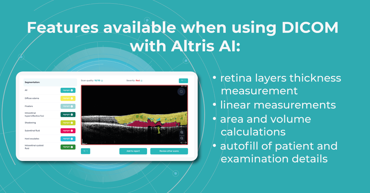
Such features as
- retina layers thickness and linear measurements
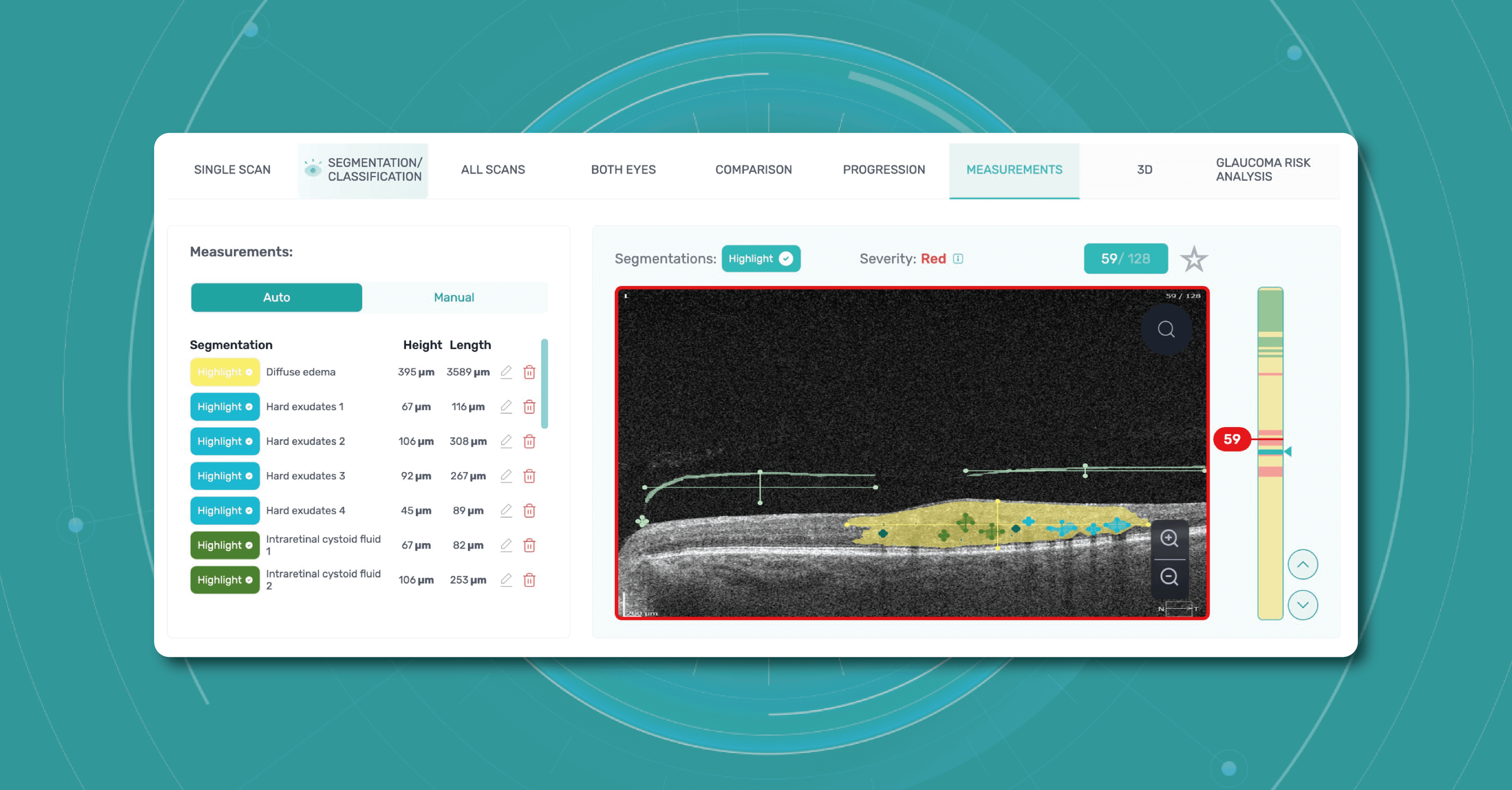
- area and volume calculations
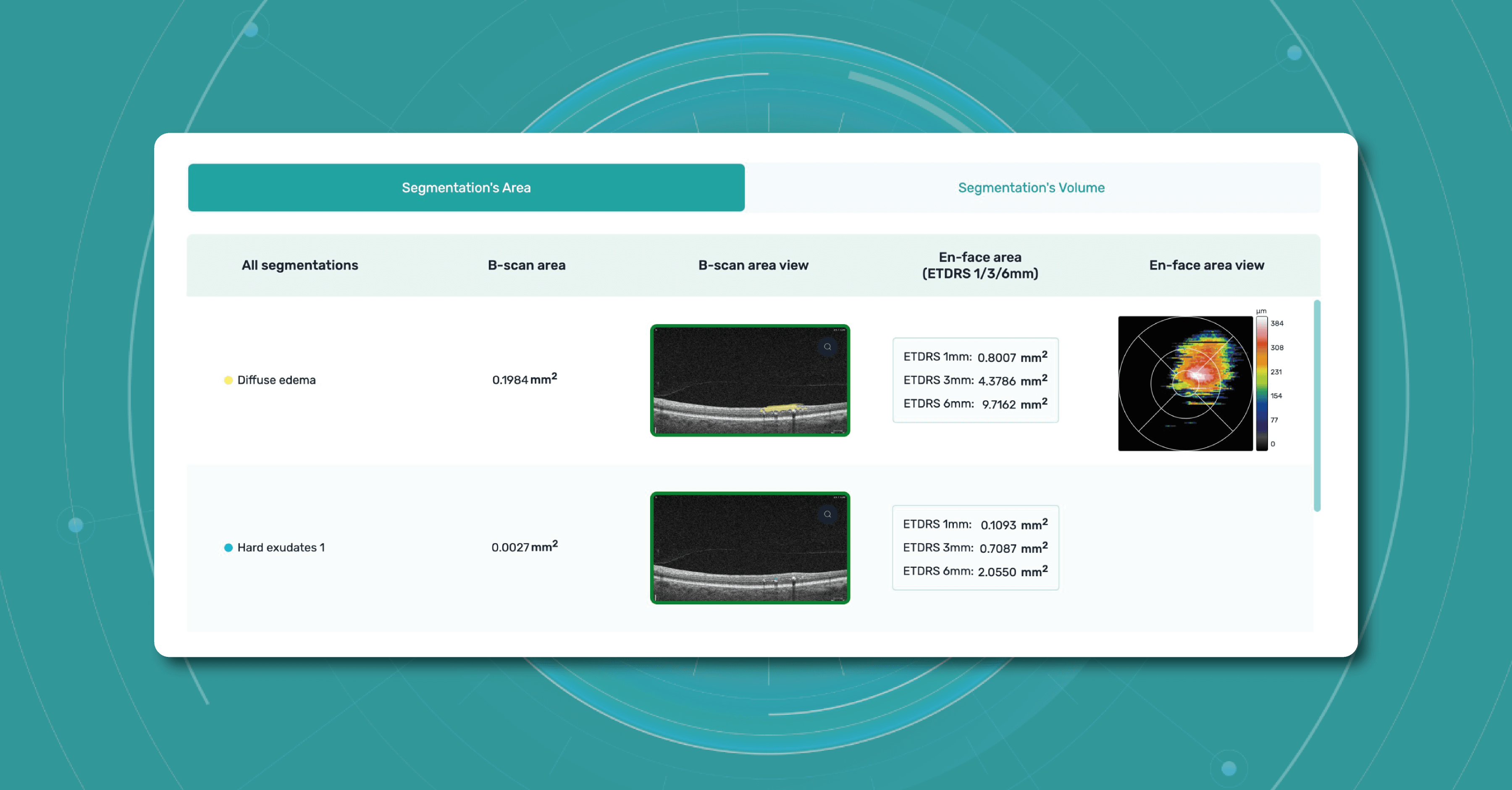
are only available when using the DICOM file format. This is because it contains the original image pixel data without modifying the study metadata. In case you upload an image, retina layers thickness won’t be available, as well as the measurements.
Another advantage of the DICOM format is that you can add patient and examination details in a few clicks by just uploading a DICOM file since this information is being pulled out automatically.
In the case of other image formats, when uploading an examination, you would have to manually fill in a bunch of information such as scan widths, eye type, etc.
Considering all mentioned above, using DICOM format files saves time, increases efficiency, and gives you more opportunities within the Altris AI platform.
Summing up
What is DICOM format? In conclusion, the DICOM file format proves to be a valuable asset for eye care specialists. Its unique characteristics, such as grouping information into data sets and incorporating standardized tags within a single file, offer many advantages.
This format ensures the preservation of accurate and comprehensive data, reducing the potential for human error and minimizing the risk of data loss or misinterpretation. The DICOM file format streamlines the archival, organization, and display of images, optimizing the workflow of eye care specialists.
By adhering to the DICOM standard, OCT devices and software solutions ensure compatibility, interoperability, and consistent data representation across different platforms. This enables efficient communication and collaboration among healthcare professionals, enhances research capabilities, and promotes the broader use and exchange of OCT imaging data.
With its widespread adoption and compatibility with various medical imaging systems, DICOM empowers ophthalmologists and optometrists to provide efficient and high-quality care while promoting seamless collaboration and knowledge sharing within the field. Ultimately, the DICOM file format plays a vital role in enhancing patient care, advancing research, and fostering innovation in the field of eye care.
FAQ
1. Why is DICOM the preferred format for ophthalmic imaging workflows?
DICOM is the industry standard because it stores both the image and full clinical metadata (patient data, scan parameters, measurements) in one structured file, enabling interoperability across devices and systems.
2. What makes DICOM better than standard image formats like JPEG or PNG in clinical use?
Unlike flat image formats, DICOM preserves diagnostic context, including acquisition settings, annotations, and structured metadata, which are essential for accurate interpretation and longitudinal tracking.
3. How does DICOM improve efficiency in AI-powered platforms like Altris AI?
DICOM allows automatic extraction of patient and scan metadata, reducing manual input and enabling faster processing, standardized workflows, and more reliable AI-based analysis of retinal images.
4. What is the key benefit of using DICOM data in AI-driven retina analysis?
It ensures full data integrity — the image pixel data and metadata remain linked, enabling precise measurements, longitudinal comparison, and more accurate AI model outputs without data loss or mismatch.