
Mark Braddon
Head of Sales
Reading time
7 min read
In recent years reading centers have become an essential resource for facilitating imaging research in many fields, including clinical trials of ophthalmology drugs. And their importance will continue to grow.
Reading centers provide crucial information by evaluating images. That is why for conducting accurate clinical trials, they must hire ophthalmologists of high qualification. Moreover, to ensure consistent analysis, the materials that graders use for the research (be it fundus photographs, fluorescein angiograms, or OCT scans) must also undergo quality control. However, even such measures can’t completely exclude errors or biases.
Meanwhile, recent developments in the field of AI medical image analysis revolutionized the approach to clinical trials, which makes it possible to boost the workflow of reading centers. AI image analysis software works with thousands of images, efficiently providing the large amount of data needed to analyze the patient’s condition. In addition, evaluating images with AI is faster, cheaper, and more effective.

Book demo for a company
Try artificial intelligence for OCT analysis
In this article, we will discuss the top 5 benefits of AI medical image analysis software for reading centers and the way AI improves the image interpretation process.
Limitations of the manual evaluating procedure
Although several reading centers have already implemented AI for medical image analysis in their workflow, most organizations are far from evaluating automation and prefer classic image interpretation methods.

In most reading centers, ophthalmologists manually evaluate ocular images for drug safety studies, compile the images, and perform statistical analysis of the data. Research sizes for reading centers can range from 50 images to 3000 or more, and dozen of separate sets of images can be collected per research subject. Therefore reading centers have many obstacles to a quality evaluation process and accurate results.
-
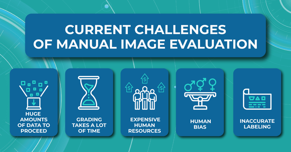
Large amount of images is hard to proceed
The vast number of images that need to be processed in the short term usually leads to the main problem for reading centers — most hire outsourced ophthalmologists to speed up the image grading and evaluation process. Outsourced specialists have different levels of qualification and different evaluating methods, which may lead to decreased accuracy. In addition, outsourced eye care specialists are not always interested in performing the work at the highest level.
-
Human resources are expensive
Another limitation of the standard evaluating procedure is the high сost spent on ophthalmologists. Human resources are usually quite expensive and associated with the risk of staff turnover.
-
High probability of human bias
Besides, hours spent in front of a computer screen evaluating thousands of images create a stressful environment for ophthalmologists and cause many errors, affecting the accuracy of the clinical trials. Even the FDA recognizes grader fatigue and its impact on potential errors in image interpretation.
-
Inaccurate labeling
In addition, administrative problems also occur quite often. This happens due to deviations from study protocols and incorrect labeling of images, which can compromise the integrity of the analyses.
Fortunately, the pace of digitalization in reading centers is accelerating. Here is how AI medical image analysis can help reading centers cope with the growing workload.
The importance of implementing AI medical image analysis for reading centers
Usually, AI image analysis is made through a pattern recognition process that involves scanning images for specific pathological signs to interpret the patient’s condition. The AI image analysis software has precise and efficient evaluation protocols that allow the analysis and interpretation of images in terms of a variety of qualitative morphological parameters. For example, when analyzing images of a patient with diabetic retinopathy, the AI models recognize microaneurysms or hemorrhages.

AI algorithms allow reading centers to conduct trials of any size and duration, including various treatments for various eye diseases. Moreover, unlike the standard image interpretation process, which requires significant human resources, the introduction of AI for image analysis into the workflow of reading centers has many advantages.
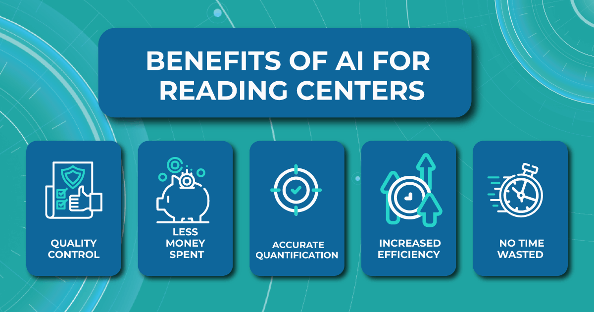
- Quality control. Using AI algorithms ensures no errors in OCT scan analysis. AI image analysis software ensures that the desired parameters are classified based on certified imaging protocols.
- Less money spent. Implementing AI-assisted OCT analysis is less expensive than hiring outsourced ophthalmologists.
- Accurate quantification. AI in medical image analysis does not depend on patient characteristics or treatment group assignment knowledge, so the machine provides the most objective and accurate assessment possible.
- Increased efficiency. Improving the reading centers workflow with AI provides an objective and standardized classification of images. It means that any human bias is excluded, which increases the reputation of clinical research.
- No time wasted — no more hours spent at a computer screen. Evaluating images with AI medical image analysis provides faster and more sensitive identification of the patient’s condition, which can positively impact decision-making.

Book demo for a company
Try artificial intelligence for OCT analysis
How reading centers will benefit from AI image analysis software
In short, image evaluation with algorithms is fast, less expensive, and more reproducible. However, many companies that perform clinical trials in cooperation with reading centers are still afraid of implementing AI in medical image analysis and evaluating processes. Modern AI-based image management systems, such as Altris AI, unlike their predecessors, allow reading centers to overcome the challenges of the manual image interpretation process.
A lot of data available to train an algorithm
The more images with various pathological features the algorithm has for training, the more accurately it will detect the diagnosis. Modern AI image analysis software has the ability to obtain thousands of OCT images from different models of devices for comprehensive and correct training of algorithms. Although many medical centers keep their clinical practice confidential, many ophthalmic cases and images with various pathological signs in the public domain allow the training of AI algorithms.
For example, the Altris AI medical image analysis software was trained on 5 million unique OCT scans obtained in 11 practicing ophthalmology clinics through the years. Our retina experts took a responsible approach to annotating and labeling images for algorithm training. A thorough error detection and correction procedure gave our algorithm 91% accuracy.
Constant quality control
The responsibilities of the modern algorithms developers include not only the release of the model but also further diagnostics, which allows avoiding the problem of reproducibility. After all, constant quality control is necessary for algorithm development environments. Understanding the importance of quality control, the Altris AI team constantly tests the reproducibility of AI medical image analysis model diagnostics.
Collection of rare diseases
According to our research, 25% of ophthalmologists, on average, miss rare pathologies 3 times a week. However, modern AI image analysis software allows overcoming this challenge. For example, Altris AI excludes missing minor, early, rare pathologies. Our team created an algorithm that automates the detection of 54 pathological signs and 49 pathologies.
High percentage of algorithmic bias is avoided
Algorithmic bias is one of the biggest challenges in AI. Although algorithms themselves do not have biases, they inherit them from humans. However, today, AI for image analysis has learned how to overcome the lack of interoperability between medical record systems.
Although it is impossible to avoid algorithmic bias completely, as it can appear at any stage of the algorithm creation process, from study design and data collection to algorithm development and model selection, modern developers take a direction to fair AI. By using a technical and regulatory framework that provides the diverse data needed to train AI algorithms, the Altris AI team makes modern technologies inclusive and ensures algorithmic bias can be excluded.
The future of AI medical image analysis in reading centers
The ultimate goal of the ophthalmic AI system for reading centers is to improve the grading and evaluating process and obtain more accurate research results. However, instead of fully digitalizing image assessment, the ideal approach to analysis is integration — where the benefits of AI algorithms and human skills can be combined.
Technology will never fully replace humans, but it is already improving their work efficiency. For example, by taking over more routine and monotonous tasks, algorithms allow ophthalmologists to focus on specific eye areas and increase the evaluation speed. AI medical image analysis software can also be effective in determining compliance with the standardization of feature interpretation and determining image quality for requesting more images.
There are undoubtedly many challenges to integrating AI for image analysis into the workflow of reading centers. However, modern AI technologies can already overcome almost all of them. Altris AI image interpretation system is changing the future of clinical research by helping to classify images faster and increasing the efficiency, accuracy, and reproducibility of clinical trial data.
You can watch a short video of how Altris AI platform assists eye care specialists in detecting pathological signs on the OCT scans:
