Featured This month
-
Inside the Power Hour: Altris AI’s Take on AI Innovation in Eye Care
 Grant Schmid
3 min
Grant Schmid
3 minInside the Power Hour: Altris AI’s Take on AI Innovation in Eye Care
Our Vice President of Business Development, Grant Schmid, took part in The Power Hour podcast to discuss how AI and automation are shaping the future of patient experience. We turned that conversation into an interview and pulled out the most compellinsubtle anatomical g insights on tech-enabled practice growth and innovation in eye care.
Eugene Shatsman: Can you start by introducing Altris AI and what problem you’re solving in eye care?
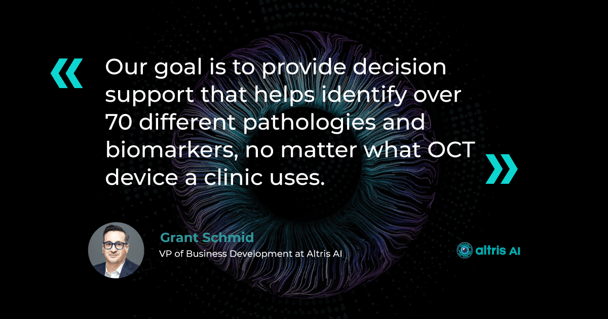
Grant Schmid: Altris AI was founded in 2017 in Chicago, with the University of Chicago as our first investor. But most of our team — and the heart of our development — is based in Ukraine.We focus on AI for OCT analysis. Our goal is to provide decision support that helps identify over 70 different pathologies and biomarkers, no matter what OCT device a clinic uses. The idea is to speed up image interpretation, ensure nothing is missed, and support doctors in delivering top-quality care.

Eugene: What initially inspired the development of Altris AI?
Grant: Our co-founder is a retina specialist from Kyiv. She wanted a way to improve the referral process and increase the OCT knowledge of those referring patients to her. That’s how the idea of a clinical decision support platform was born.We actually started with an educational OCT app that you can still download — many doctors come to our booth at trade shows not realizing that the app is also part of what we’ve built.
Eugene: What does a typical OCT workflow look like with and without Altris AI?
Grant: In many modern practices, every patient now gets an OCT. It’s used to screen for diseases like AMD, glaucoma, or diabetic retinopathy. But subtle anatomical differences can confuse even experienced clinicians.
Learn more about Altris AI’s Decision Support for OCT analysis
With Altris AI, the doctor gets an analysis almost immediately — color-coded overlays, pathology markers, optic disc assessments, all in one place. This speeds up the review process and supports clinical decision-making without disrupting workflow.
Eugene: What do you say to clinicians who say, “I already know how to read OCTs — why do I need AI?”
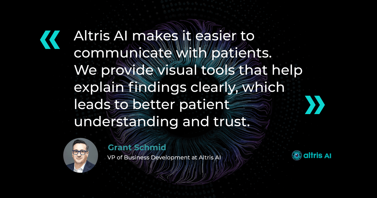
Grant: Many doctors are confident in interpreting OCTs, and that’s great. But the value isn’t just in identifying disease — it’s in validation and patient education.We’re not here to replace what doctors do. Altris AI validates what you already know and makes it easier to communicate with patients. We highlight what might be missed, and we provide visual tools that help explain findings clearly — which leads to better patient understanding and trust.
Eugene: Can you give an example of how this helps patient education?
Grant: Absolutely. Let’s take glaucoma. Many patients on drops don’t feel or see any change, so they think, “Why bother?” But if you can show them a progression or show that things are stable, it becomes real to them.We launched an Optic Disc Analysis feature that lets you compare up to eight past visits side-by-side. So when a patient asks, “Is this working?” you can say, “Yes, here’s the proof.” That drives adherence and builds trust.
Eugene: Are practices today ready to embrace AI-based tools? Or are they still cautious?
Grant: There’s a lot of curiosity, a lot of interest. Some are still figuring out how to implement AI in a way that makes sense for them.But AI is everywhere now — whether it’s in search engines, smartphones, or how we shop. Patients expect that kind of intelligence in their healthcare, too. In fact, a 67-year-old tugboat captain with AMD once called me asking about our software and offered to pay for his doctor’s subscription. That tells you how fast expectations are changing.
Eugene: Can AI actually improve the patient experience beyond just diagnosis?
Grant: Absolutely. Patients want to understand what’s happening with their health. When you can show them their scan results with overlays and simple visuals, they feel included in the process.It’s not just about detecting disease, it’s about building trust. Clear visual communication boosts confidence, reduces anxiety, and increases compliance.
Learn more about Altris AI’s Decision Support for OCT analysis
Eugene: Some fear AI will replace clinicians. What’s your perspective on that?
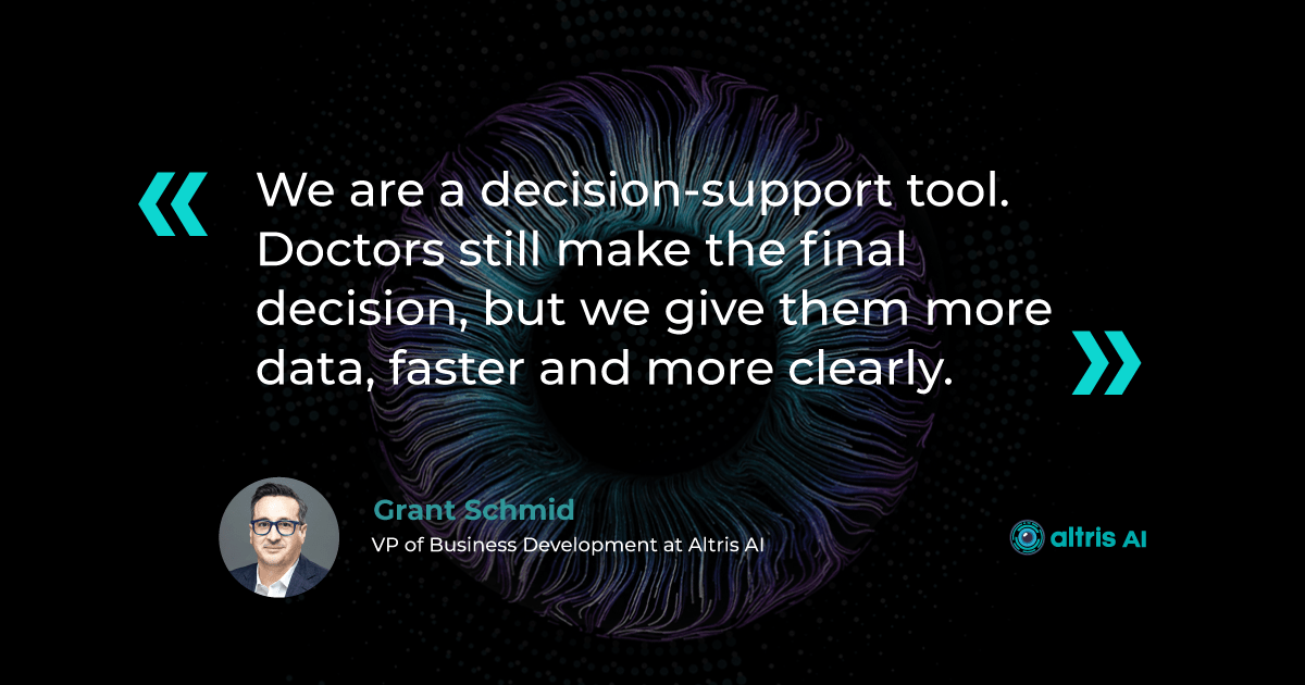
Grant: That’s one of the biggest myths out there. AI won’t replace clinicians — it enhances what they do.We’re not cleared to diagnose. We’re a decision-support tool. Doctors still make the final decision, but we give them more data, faster and more clearly. Human clinical judgment is still irreplaceable — we just help sharpen it.
Eugene: What barriers are you seeing when introducing Altris AI to new practices?
Grant: The main one is comfort — many doctors feel confident reading OCTs and don’t immediately see the need.The other is simply awareness. We’re a fast-growing startup, but many still don’t know about us. That’s why opportunities like this podcast are important.
In terms of logistics, there’s no barrier. Altris AI is web-based, nothing to install, and takes just 20 minutes to learn. We’re designed to be plug-and-play.
Eugene: If a practice wants to engage patients more using AI in eye care, how should they approach it?
Grant: One great idea is to run a recall campaign for patients who haven’t had an OCT in the last 6 or 12 months. Something like, “We now use AI to enhance your OCT scan — come see how it works.”AI is a differentiator. It shows your clinic is modern, patient-focused, and using the best available tools.
Eugene: What do you think the optometry practice of 2028 will look like?
Grant: I think you’ll see AI systems talking to each other. Imagine our platform detecting something on a scan and automatically triggering a patient reminder or a suggested follow-up.There will be less manual work and more focus on human care. The doctor will be able to walk in and focus completely on the patient — the AI will handle the background tasks like charting or longitudinal comparisons.
Ultimately, better care, less burnout.
Eugene: What’s one myth you’d like to bust about AI in optometry?
Grant: That AI will replace people. It won’t. What it does is make you more effective. You’ll have sharper insights, clearer visuals, and faster decision-making — all without replacing your clinical experience.Eugene: And finally, how can practices get started with Altris AI?
Grant: Just go to altris.ai or connect with us on LinkedIn. We offer live demos and can use your real OCT scans to show exactly how it works.There’s no software to install, no major investment, and we operate on a subscription basis — so there’s no long-term risk. If you’re curious, reach out. We’d love to show you what’s possible.
Watch the complete Power Hour podcast episode below for more insights on AI, automation, and innovation in eye care:
-
Dry AMD Treatment: How to Slow Progression with Modern Approaches
 Maria Znamenska
5 min.
Maria Znamenska
5 min.Dry AMD Treatment: How to Slow Progression with Modern Approaches
Table of Contents
1.What are the dry macular degeneration treatment breakthroughs?
2.How to monitor dry AMD progression with OCT?
3.What are the challenges of dry age-related macular degeneration monitoring?
4.How do I organize efficient dry AMD monitoring in my clinic?
5.Why are optometrists on the front line of early AMD detection?
6.How can OCT insights help support patients emotionally?
For many years, dry or non-exudative AMD was considered untreatable. Most efforts were focused on treating the wet or exudative AMD with anti-VEGF drugs. However, this paradigm has recently shifted.
The first FDA-approved drugs appeared recently to treat geographic atrophy (GA), which affects 30% of patients with dry AMD. Additionally, new physiotherapeutic methods, such as multi-wavelength photobiomodulation, have emerged.
Geographic atrophy (GA) is an advanced, irreversible form of dry age-related macular degeneration (AMD). It develops when areas of the retina, the light-sensitive tissue at the back of the eye, undergo cell death (atrophy), causing progressive vision loss.
However, even the best dry AMD treatment is ineffective without an objective way to measure its success. Updated guidelines suggest advanced tools for monitoring progression, and optical coherence tomography (OCT) is at the core of this process.
What are the dry macular degeneration treatment breakthroughs?
The dry macular degeneration treatment breakthroughs include multiwavelength photobiomodulation, FDA-approved injectable drugs, and AREDS 2-based supplements. Unlike older recommendations focused on reducing risk factors — quitting smoking, managing blood pressure, and eating a healthy diet — these new approaches for dry AMD combine prevention with active treatment strategies to slow the progression of GA.
1. Dry AMD treatment using multiwavelength photobiomodulation
Multiwavelength photobiomodulation for AMD is a promising new treatment. It uses specific light wavelengths (in the red and near-infrared spectrum, ~590 to 850 nm) to reduce oxidative stress, inflammation, and pigment epithelial cell death in the retina.
One of the most well-known systems used for this approach is Valeda Light Therapy, which delivers controlled multiwavelength light to the retina in a non-invasive manner.
The LIGHTSITE III clinical trial (2022) showed that photobiomodulation significantly slowed the decline in visual acuity and reduced the rate of GA expansion.
Limitations:
- Limited long-term data (only 3–5 years available)
- Requires expensive equipment and trained personnel
- Unclear effectiveness in late-stage GA

2. Dry AMD treatment using FDA-approved injectable drugs
AMD injection drugs approved by the FDA include Izervay and Syfovre.
- Izervay (avacincaptad pegol): A C5 complement protein inhibitor that targets the complement cascade involved in chronic retinal inflammation and damage. Izervay, approved for geographic atrophy secondary to dry AMD, has demonstrated a reduced rate of GA progression in clinical trials.
- Syfovre (pegcetacoplan): A C3 complement inhibitor that blocks the central component of the complement system to reduce inflammation. Syfovre is the first FDA-approved treatment for GA that targets complement component C3, showing a clinically meaningful slowing of GA progression.
Both dry macular degeneration injections have shown the ability to slow GA progression compared to placebo. Although they do not restore vision, slowing vision loss is a meaningful clinical outcome.
Usage considerations:
- Administered via intravitreal injections, usually monthly or every other month
- Doctors need training; patients must be informed about risks (e.g., endophthalmitis, increased IOP)
- Cost and availability may be barriers
3. Dry AMD treatment using AREDS 2-based supplements
AREDS 2 supplements are antioxidant supplements containing lutein, zeaxanthin, vitamins C and E, zinc, and copper. They can reduce the risk of progression to late stage AMD by around 25% over five years, according to the AREDS 2 study.
Pros:
- Easily accessible
- Low risk of side effects
- A strong evidence base
Cons:
- Does not directly affect GA
- Cannot replace active treatments like injections or photobiomodulation
How to monitor dry AMD progression with OCT?
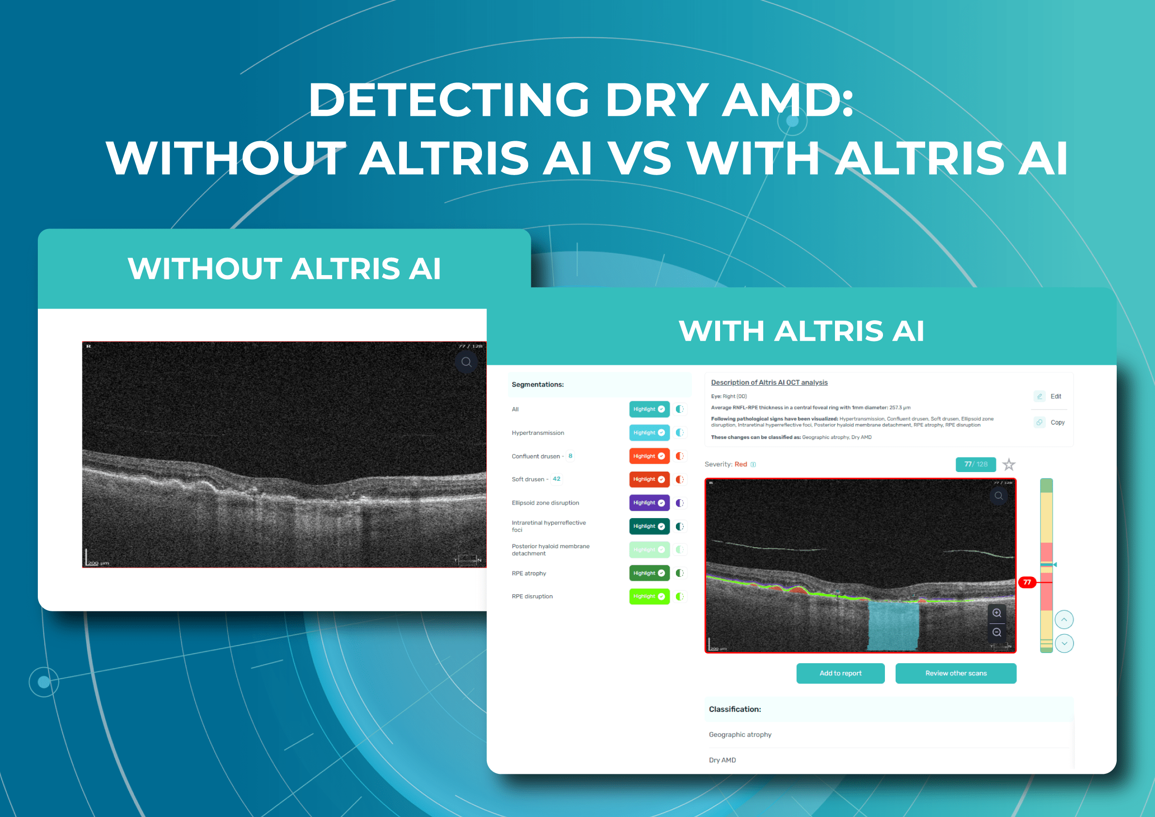
To monitor dry AMD progression effectively, OCT is essential. It is the gold standard for tracking structural changes in the retina. Without OCT, clinicians are essentially flying blind when it comes to assessing disease progression and predicting geographic atrophy (GA) development.
What are the key monitoring parameters of AMD progression?
The key monitoring parameters of AMD progression include GA area, drusen, and distance to fovea.
1. GA area
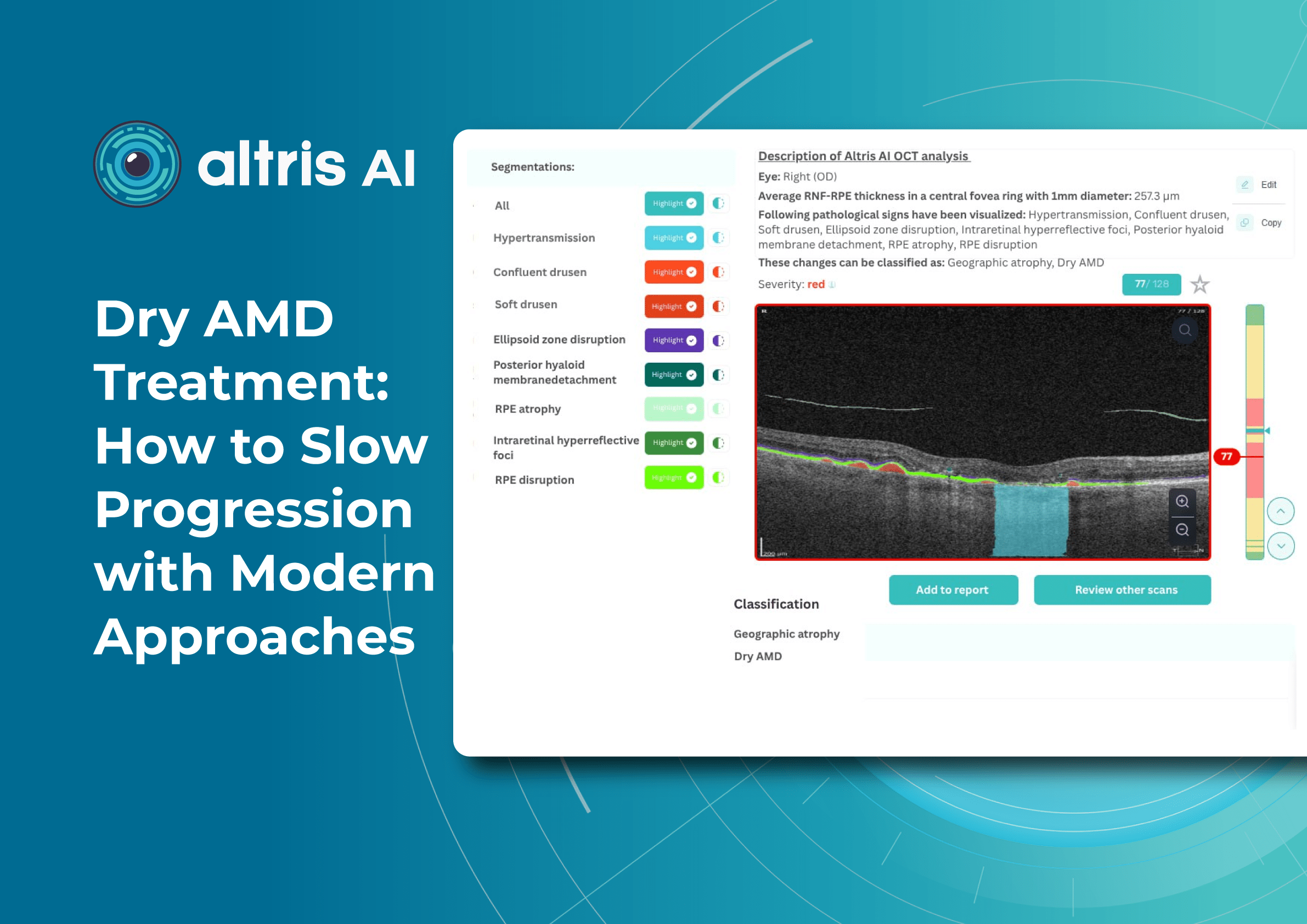
This is the main metric when using intravitreal eye injections. Modern OCT systems provide GA measurements in mm², allowing doctors to objectively track changes over time.
Even if patients don’t notice symptoms, a growing GA area signals disease progression. In FDA trials for Syfovre and Izervay, the GA area was the primary endpoint.
2. Drusen
Drusen vary in number, size, and shape. A reduction or disappearance of drusen on OCT may seem like an improvement, but could actually indicate a transition to the atrophic stage. Regular monitoring helps detect this early.
3. Distance to fovea
The closer GA is to the fovea, the greater the risk of sudden vision loss.
Early detection enables:
- Referral to an ophthalmologist
- Timely conversations about potential vision loss
What are OCT outputs for AMD progression monitoring and communication?
Useful OCT outputs for AMD progression monitoring and communication are heat maps and progress charts.
1. Heat maps
Modern OCT systems use color-coded heat maps to show pigment epithelium thickness and drusen distribution. This visual format helps in several ways:
- Makes interpretation easier for clinicians
- Helps patients better understand their condition
- Encourages patients to stay engaged with treatment
In clinical practice, it serves as a highly effective communication tool.
2. Progress charts
Most OCT systems can compare results across visits
- For doctors: Helps guide treatment decisions
- For patients: Provides visual proof of stabilization or worsening
The role of objective evidence in patient treatment
Patients may question the value of long-term treatments or costly procedures.
OCT is the gold standard for patient motivation. When patients see actual changes, they’re more likely to agree to treatment.
What are the challenges of dry age-related macular degeneration monitoring?
Monitoring dry AMD presents technical, organizational, and psychological challenges. Doctors of all levels of experience should be aware of them.
1. Invisible microchanges
Early atrophy or drusen changes may be subtle. Patients may not notice them due to eccentric fixation or slow adaptation.
Without OCT, doctors may miss early GA, delaying treatment.
It is necessary to perform OCT even when there are only minor changes in visual acuity or if the patient reports image distortion (metamorphopsia).
2. Subjective assessment
Ophthalmoscopy reveals only obvious changes. Subtle drusen or early atrophy might be missed.
Relying on patients’ complaints is risky — many don’t notice issues until it’s too late.
That’s why even small optical practices should establish clear referral pathways for OCT exams.
3. Unnecessary referrals
Optometrists or primary care doctors often refer patients to ophthalmologists “just in case,” because they don’t have access to OCT or lack experience interpreting it.
This puts unnecessary strain on specialists. In many cases, nothing new is done after the exam because there are no previous images for comparison.
4. Limitations of OCT devices
Not all OCT devices measure GA or track drusen equally well. Older models may lack automated measurements of atrophy area.
In some cases, referral to a center with advanced OCT is necessary.
How do I organize efficient dry AMD monitoring in my clinic?
Here’s how you can organize efficient monitoring in your clinic:
Tip 1. Create a baseline chart
During the first visit, perform a detailed OCT scan to measure GA area, evaluate drusen, and record distance to the fovea. Save the images or print them for future comparison.
Tip 2. Monitor frequently
- Early stages: every 6–12 months
- With GA: every 3–6 months
- When treated with intravitreal injections: before each injection
A reminder system helps with patient compliance.
Tip 3. Standardize your protocol
Use the same scanning protocols every time to reduce variability.
Tip 4. Use OCT software tools
Modern systems offer:
- Image comparison
- Automatic GA area calculation
- Heat map visualization
Tip 5. Communicate clearly with patients
Use simple language:
- These are areas of atrophy, and we’re measuring them
- These bright spots are drusen we’re monitoring
- The goal is to slow the growth of these areas
Educated patients are more engaged in their care.
Why are optometrists on the front line of early AMD detection?
Optometrists play a key role in spotting the early signs of AMD, as they are often the first point of contact in eye care.
They perform initial screenings, provide guidance on lifestyle and supplements, and ensure regular OCT monitoring.
If drusen, pigment epithelial changes, or signs of GA are present, they refer patients to ophthalmologists for confirmation and treatment planning.
How can OCT insights help support patients emotionally?
Explaining a chronic, progressive condition like AMD to elderly patients can be difficult. Motivating them to return for regular follow-ups is often even harder.
Many ask, “Why bother if it can’t be cured?”
OCT insights can support both understanding and emotional reassurance. A thoughtful approach may include:
-
Explaining that treatment helps slow vision loss
-
Emphasising their active role in preserving sight
-
Using OCT scans to show visual proof of stability or progress
Conclusion
Modern dry AMD treatment is no longer a dead end. With FDA-approved medications, photobiomodulation, and effective supplements, optometrists can significantly impact disease progression.
But none of this works without quality monitoring. OCT reveals what the eye can’t see and helps guide clinical decisions while motivating patients.
The ultimate goal is to partner with patients in preserving their vision. This isn’t a one-time visit—it’s a long-term commitment. The stronger the support, the better the chances of maintaining central vision and seeing meaningful results from dry AMD treatment.


popular Posted
-
Inside the Power Hour: Altris AI’s Take on AI Innovation in Eye Care
 Grant Schmid
3 min
Grant Schmid
3 minInside the Power Hour: Altris AI’s Take on AI Innovation in Eye Care
Our Vice President of Business Development, Grant Schmid, took part in The Power Hour podcast to discuss how AI and automation are shaping the future of patient experience. We turned that conversation into an interview and pulled out the most compellinsubtle anatomical g insights on tech-enabled practice growth and innovation in eye care.
Eugene Shatsman: Can you start by introducing Altris AI and what problem you’re solving in eye care?
Grant Schmid: Altris AI was founded in 2017 in Chicago, with the University of Chicago as our first investor. But most of our team — and the heart of our development — is based in Ukraine.We focus on AI for OCT analysis. Our goal is to provide decision support that helps identify over 70 different pathologies and biomarkers, no matter what OCT device a clinic uses. The idea is to speed up image interpretation, ensure nothing is missed, and support doctors in delivering top-quality care.
Eugene: What initially inspired the development of Altris AI?
Grant: Our co-founder is a retina specialist from Kyiv. She wanted a way to improve the referral process and increase the OCT knowledge of those referring patients to her. That’s how the idea of a clinical decision support platform was born.We actually started with an educational OCT app that you can still download — many doctors come to our booth at trade shows not realizing that the app is also part of what we’ve built.
Eugene: What does a typical OCT workflow look like with and without Altris AI?
Grant: In many modern practices, every patient now gets an OCT. It’s used to screen for diseases like AMD, glaucoma, or diabetic retinopathy. But subtle anatomical differences can confuse even experienced clinicians.
Learn more about Altris AI’s Decision Support for OCT analysis
With Altris AI, the doctor gets an analysis almost immediately — color-coded overlays, pathology markers, optic disc assessments, all in one place. This speeds up the review process and supports clinical decision-making without disrupting workflow.
Eugene: What do you say to clinicians who say, “I already know how to read OCTs — why do I need AI?”
Grant: Many doctors are confident in interpreting OCTs, and that’s great. But the value isn’t just in identifying disease — it’s in validation and patient education.We’re not here to replace what doctors do. Altris AI validates what you already know and makes it easier to communicate with patients. We highlight what might be missed, and we provide visual tools that help explain findings clearly — which leads to better patient understanding and trust.
Eugene: Can you give an example of how this helps patient education?
Grant: Absolutely. Let’s take glaucoma. Many patients on drops don’t feel or see any change, so they think, “Why bother?” But if you can show them a progression or show that things are stable, it becomes real to them.We launched an Optic Disc Analysis feature that lets you compare up to eight past visits side-by-side. So when a patient asks, “Is this working?” you can say, “Yes, here’s the proof.” That drives adherence and builds trust.
Eugene: Are practices today ready to embrace AI-based tools? Or are they still cautious?
Grant: There’s a lot of curiosity, a lot of interest. Some are still figuring out how to implement AI in a way that makes sense for them.But AI is everywhere now — whether it’s in search engines, smartphones, or how we shop. Patients expect that kind of intelligence in their healthcare, too. In fact, a 67-year-old tugboat captain with AMD once called me asking about our software and offered to pay for his doctor’s subscription. That tells you how fast expectations are changing.
Eugene: Can AI actually improve the patient experience beyond just diagnosis?
Grant: Absolutely. Patients want to understand what’s happening with their health. When you can show them their scan results with overlays and simple visuals, they feel included in the process.It’s not just about detecting disease, it’s about building trust. Clear visual communication boosts confidence, reduces anxiety, and increases compliance.
Learn more about Altris AI’s Decision Support for OCT analysis
Eugene: Some fear AI will replace clinicians. What’s your perspective on that?
Grant: That’s one of the biggest myths out there. AI won’t replace clinicians — it enhances what they do.We’re not cleared to diagnose. We’re a decision-support tool. Doctors still make the final decision, but we give them more data, faster and more clearly. Human clinical judgment is still irreplaceable — we just help sharpen it.
Eugene: What barriers are you seeing when introducing Altris AI to new practices?
Grant: The main one is comfort — many doctors feel confident reading OCTs and don’t immediately see the need.The other is simply awareness. We’re a fast-growing startup, but many still don’t know about us. That’s why opportunities like this podcast are important.
In terms of logistics, there’s no barrier. Altris AI is web-based, nothing to install, and takes just 20 minutes to learn. We’re designed to be plug-and-play.
Eugene: If a practice wants to engage patients more using AI in eye care, how should they approach it?
Grant: One great idea is to run a recall campaign for patients who haven’t had an OCT in the last 6 or 12 months. Something like, “We now use AI to enhance your OCT scan — come see how it works.”AI is a differentiator. It shows your clinic is modern, patient-focused, and using the best available tools.
Eugene: What do you think the optometry practice of 2028 will look like?
Grant: I think you’ll see AI systems talking to each other. Imagine our platform detecting something on a scan and automatically triggering a patient reminder or a suggested follow-up.There will be less manual work and more focus on human care. The doctor will be able to walk in and focus completely on the patient — the AI will handle the background tasks like charting or longitudinal comparisons.
Ultimately, better care, less burnout.
Eugene: What’s one myth you’d like to bust about AI in optometry?
Grant: That AI will replace people. It won’t. What it does is make you more effective. You’ll have sharper insights, clearer visuals, and faster decision-making — all without replacing your clinical experience.Eugene: And finally, how can practices get started with Altris AI?
Grant: Just go to altris.ai or connect with us on LinkedIn. We offer live demos and can use your real OCT scans to show exactly how it works.There’s no software to install, no major investment, and we operate on a subscription basis — so there’s no long-term risk. If you’re curious, reach out. We’d love to show you what’s possible.
Watch the complete Power Hour podcast episode below for more insights on AI, automation, and innovation in eye care:
-
Dry AMD Treatment: How to Slow Progression with Modern Approaches
Maria Znamenska
5 min.Dry AMD Treatment: How to Slow Progression with Modern Approaches
Table of Contents
1.What are the dry macular degeneration treatment breakthroughs?
2.How to monitor dry AMD progression with OCT?
3.What are the challenges of dry age-related macular degeneration monitoring?
4.How do I organize efficient dry AMD monitoring in my clinic?
5.Why are optometrists on the front line of early AMD detection?
6.How can OCT insights help support patients emotionally?
For many years, dry or non-exudative AMD was considered untreatable. Most efforts were focused on treating the wet or exudative AMD with anti-VEGF drugs. However, this paradigm has recently shifted.
The first FDA-approved drugs appeared recently to treat geographic atrophy (GA), which affects 30% of patients with dry AMD. Additionally, new physiotherapeutic methods, such as multi-wavelength photobiomodulation, have emerged.
Geographic atrophy (GA) is an advanced, irreversible form of dry age-related macular degeneration (AMD). It develops when areas of the retina, the light-sensitive tissue at the back of the eye, undergo cell death (atrophy), causing progressive vision loss.
However, even the best dry AMD treatment is ineffective without an objective way to measure its success. Updated guidelines suggest advanced tools for monitoring progression, and optical coherence tomography (OCT) is at the core of this process.
What are the dry macular degeneration treatment breakthroughs?
The dry macular degeneration treatment breakthroughs include multiwavelength photobiomodulation, FDA-approved injectable drugs, and AREDS 2-based supplements. Unlike older recommendations focused on reducing risk factors — quitting smoking, managing blood pressure, and eating a healthy diet — these new approaches for dry AMD combine prevention with active treatment strategies to slow the progression of GA.
1. Dry AMD treatment using multiwavelength photobiomodulation
Multiwavelength photobiomodulation for AMD is a promising new treatment. It uses specific light wavelengths (in the red and near-infrared spectrum, ~590 to 850 nm) to reduce oxidative stress, inflammation, and pigment epithelial cell death in the retina.
One of the most well-known systems used for this approach is Valeda Light Therapy, which delivers controlled multiwavelength light to the retina in a non-invasive manner.
The LIGHTSITE III clinical trial (2022) showed that photobiomodulation significantly slowed the decline in visual acuity and reduced the rate of GA expansion.
Limitations:
- Limited long-term data (only 3–5 years available)
- Requires expensive equipment and trained personnel
- Unclear effectiveness in late-stage GA
2. Dry AMD treatment using FDA-approved injectable drugs
AMD injection drugs approved by the FDA include Izervay and Syfovre.
- Izervay (avacincaptad pegol): A C5 complement protein inhibitor that targets the complement cascade involved in chronic retinal inflammation and damage. Izervay, approved for geographic atrophy secondary to dry AMD, has demonstrated a reduced rate of GA progression in clinical trials.
- Syfovre (pegcetacoplan): A C3 complement inhibitor that blocks the central component of the complement system to reduce inflammation. Syfovre is the first FDA-approved treatment for GA that targets complement component C3, showing a clinically meaningful slowing of GA progression.
Both dry macular degeneration injections have shown the ability to slow GA progression compared to placebo. Although they do not restore vision, slowing vision loss is a meaningful clinical outcome.
Usage considerations:
- Administered via intravitreal injections, usually monthly or every other month
- Doctors need training; patients must be informed about risks (e.g., endophthalmitis, increased IOP)
- Cost and availability may be barriers
3. Dry AMD treatment using AREDS 2-based supplements
AREDS 2 supplements are antioxidant supplements containing lutein, zeaxanthin, vitamins C and E, zinc, and copper. They can reduce the risk of progression to late stage AMD by around 25% over five years, according to the AREDS 2 study.
Pros:
- Easily accessible
- Low risk of side effects
- A strong evidence base
Cons:
- Does not directly affect GA
- Cannot replace active treatments like injections or photobiomodulation
How to monitor dry AMD progression with OCT?
To monitor dry AMD progression effectively, OCT is essential. It is the gold standard for tracking structural changes in the retina. Without OCT, clinicians are essentially flying blind when it comes to assessing disease progression and predicting geographic atrophy (GA) development.
What are the key monitoring parameters of AMD progression?
The key monitoring parameters of AMD progression include GA area, drusen, and distance to fovea.
1. GA area
This is the main metric when using intravitreal eye injections. Modern OCT systems provide GA measurements in mm², allowing doctors to objectively track changes over time.
Even if patients don’t notice symptoms, a growing GA area signals disease progression. In FDA trials for Syfovre and Izervay, the GA area was the primary endpoint.
2. Drusen
Drusen vary in number, size, and shape. A reduction or disappearance of drusen on OCT may seem like an improvement, but could actually indicate a transition to the atrophic stage. Regular monitoring helps detect this early.
3. Distance to fovea
The closer GA is to the fovea, the greater the risk of sudden vision loss.
Early detection enables:
- Referral to an ophthalmologist
- Timely conversations about potential vision loss
What are OCT outputs for AMD progression monitoring and communication?
Useful OCT outputs for AMD progression monitoring and communication are heat maps and progress charts.
1. Heat maps
Modern OCT systems use color-coded heat maps to show pigment epithelium thickness and drusen distribution. This visual format helps in several ways:
- Makes interpretation easier for clinicians
- Helps patients better understand their condition
- Encourages patients to stay engaged with treatment
In clinical practice, it serves as a highly effective communication tool.
2. Progress charts
Most OCT systems can compare results across visits
- For doctors: Helps guide treatment decisions
- For patients: Provides visual proof of stabilization or worsening
The role of objective evidence in patient treatment
Patients may question the value of long-term treatments or costly procedures.
OCT is the gold standard for patient motivation. When patients see actual changes, they’re more likely to agree to treatment.
What are the challenges of dry age-related macular degeneration monitoring?
Monitoring dry AMD presents technical, organizational, and psychological challenges. Doctors of all levels of experience should be aware of them.
1. Invisible microchanges
Early atrophy or drusen changes may be subtle. Patients may not notice them due to eccentric fixation or slow adaptation.
Without OCT, doctors may miss early GA, delaying treatment.
It is necessary to perform OCT even when there are only minor changes in visual acuity or if the patient reports image distortion (metamorphopsia).
2. Subjective assessment
Ophthalmoscopy reveals only obvious changes. Subtle drusen or early atrophy might be missed.
Relying on patients’ complaints is risky — many don’t notice issues until it’s too late.
That’s why even small optical practices should establish clear referral pathways for OCT exams.
3. Unnecessary referrals
Optometrists or primary care doctors often refer patients to ophthalmologists “just in case,” because they don’t have access to OCT or lack experience interpreting it.
This puts unnecessary strain on specialists. In many cases, nothing new is done after the exam because there are no previous images for comparison.
4. Limitations of OCT devices
Not all OCT devices measure GA or track drusen equally well. Older models may lack automated measurements of atrophy area.
In some cases, referral to a center with advanced OCT is necessary.
How do I organize efficient dry AMD monitoring in my clinic?
Here’s how you can organize efficient monitoring in your clinic:
Tip 1. Create a baseline chart
During the first visit, perform a detailed OCT scan to measure GA area, evaluate drusen, and record distance to the fovea. Save the images or print them for future comparison.
Tip 2. Monitor frequently
- Early stages: every 6–12 months
- With GA: every 3–6 months
- When treated with intravitreal injections: before each injection
A reminder system helps with patient compliance.
Tip 3. Standardize your protocol
Use the same scanning protocols every time to reduce variability.
Tip 4. Use OCT software tools
Modern systems offer:
- Image comparison
- Automatic GA area calculation
- Heat map visualization
Tip 5. Communicate clearly with patients
Use simple language:
- These are areas of atrophy, and we’re measuring them
- These bright spots are drusen we’re monitoring
- The goal is to slow the growth of these areas
Educated patients are more engaged in their care.
Why are optometrists on the front line of early AMD detection?
Optometrists play a key role in spotting the early signs of AMD, as they are often the first point of contact in eye care.
They perform initial screenings, provide guidance on lifestyle and supplements, and ensure regular OCT monitoring.
If drusen, pigment epithelial changes, or signs of GA are present, they refer patients to ophthalmologists for confirmation and treatment planning.
How can OCT insights help support patients emotionally?
Explaining a chronic, progressive condition like AMD to elderly patients can be difficult. Motivating them to return for regular follow-ups is often even harder.
Many ask, “Why bother if it can’t be cured?”
OCT insights can support both understanding and emotional reassurance. A thoughtful approach may include:
-
Explaining that treatment helps slow vision loss
-
Emphasising their active role in preserving sight
-
Using OCT scans to show visual proof of stability or progress
Conclusion
Modern dry AMD treatment is no longer a dead end. With FDA-approved medications, photobiomodulation, and effective supplements, optometrists can significantly impact disease progression.
But none of this works without quality monitoring. OCT reveals what the eye can’t see and helps guide clinical decisions while motivating patients.
The ultimate goal is to partner with patients in preserving their vision. This isn’t a one-time visit—it’s a long-term commitment. The stronger the support, the better the chances of maintaining central vision and seeing meaningful results from dry AMD treatment.
-
AItris AI for Buchanan Optometrists
 Mark Braddon
3 min.
Mark Braddon
3 min.Buchanan Optometrists and Audiologists is no ordinary eye-care center.
The Association of Optometrists (AOP) estimates 17,500 registered optometrists working across roughly 6,000 practices in the UK. The UK Optician Awards recognise the best in the UK Optical industry. To even make the top 5 is our equivalent of an Oscar nomination! They are the only practice in the UK to consistently make the top 5 since 2008. Buchanan Optometrists describe themselves as innovators who “continually push boundaries.”
Their list of awards speaks for itself:
- 2012 – National Optician Award for Premium Lens Practice of the Year
- 2013 – Luxury Eyewear Retailer of the Year and Premium Lens Practice of the Year
- 2013 – Winner at the UK Optician Awards
- 2015–2016 – Best UK Independent Practice
- 2017–2018 – Optometrist of the Year, with Alisdair Buchanan named the top optometrist in the UK
- 2023–2024 – Best Independent Optician and Best Technology Practice
And this list is not finished, as Alisdair Buchanan, the Owner and the Director of the center, is investing in their growth continuously.

With a track record like this, it’s no surprise that Buchanan Optometrists was among the first to adopt AI for Decision Support in OCT. AI is rapidly becoming a vital part of modern eye care, and leading centers are already embracing it.
Mark Braddon, Altris AI VP of Clinical Sales, sat down with Alisdair Buchanan, the owner and director of the practice, to talk about his experience with AI and what it means for the future of optometry.
Mark Braddon: You’ve been working with OCT for years. What changed in your practice after bringing in Altris AI Decision Support for OCT?
Alisdair Buchanan, Owner: As someone already confident in interpreting scans, I didn’t need help understanding OCT—but Altris provides something even more valuable: a kind of second opinion. It supports my clinical decisions and offers an added layer of reassurance, particularly in borderline or complex cases. That’s not just helpful—it’s powerful.
I didn’t think our OCT assessments could improve much—until we started using Altris AI. It’s not just an upgrade; it’s become an indispensable part of delivering modern, high-quality eye care. Altris AI has significantly enhanced the way we interpret OCT scans. What used to require prolonged focus and cross-referencing now takes moments, without sacrificing accuracy or depth. The system analyses images with incredible precision, highlighting subtle pathological changes that are often time-consuming to detect, especially during a busy clinic day.
Mark Braddon: What was the first real benefit you noticed after bringing Altris AI into your day-to-day routine?
Alisdair Buchanan, Owner: One of the most immediate benefits has been in patient communication. The platform generates clear, colour-coded visuals that make explaining findings effortless. Instead of trying to talk patients through grainy greyscale images, we can now show them precisely what we’re seeing. It’s improved understanding, reduced anxiety, and increased trust in the care we’re providing.
Mark Braddon: Was it easy to fit AI Decision Support into your OCT workflow? How easy did you find integrating Altris AI?
Alisdair Buchanan, Owner: Integration was seamless—no faff, no friction. It fits naturally into our existing workflow, with scans uploaded and analysed within seconds. It’s helped us work more efficiently, without compromising the thoroughness our patients expect.
In short, Altris AI has sharpened our clinical edge and strengthened the service we offer. It doesn’t replace experience—it enhances it. And that, for me, is the real value.
Mark Braddon: In your experience, where has AI been the most helpful in clinical work?
Alisdair Buchanan, Owner: The main area where it shines is in picking up early macular changes, particularly dry AMD. Things like drusen or subtle changes in the outer retinal layers, which could easily be missed at a glance, are brought to the surface immediately.
It’s also been handy with diabetic patients. Just having that extra layer of input to flag microstructural changes helps us stay ahead of progression.
We’ve also started using it with glaucoma suspects. While our Heidelberg Spectralis remains our go-to for structural monitoring, having the RNFL analysis from Altris adds a checkpoint. I’d never base a referral purely on it, but it’s nice to have a second opinion—even if it’s an AI one.
Mark Braddon: Has AI Decision Support changed how you handle borderline or difficult-to-call cases?
Alisdair Buchanan, Owner: I’d say it’s given us more confidence, particularly in the grey areas—those borderline cases where you’re not quite sure if it’s time to refer or just monitor a bit more closely. With AMD, for example, it has helped us catch early signs of progression and refer patients before things become urgent.
And for glaucoma, again, it’s not replacing anything we do—it’s just another tool we can lean on. Sometimes it confirms what we already thought, and other times it nudges us to look again more carefully.
Mark Braddon: How has using AI impacted your conversations with patients during consultations?
Alisdair Buchanan, Owner: One of the unexpected benefits has been how much it helps with patient conversations. We show the scans on-screen during the consultation, and the colour overlays make things much easier to explain, especially with older patients. They can see what we’re talking about, which makes the whole thing feel more real and less abstract.
They often say, “Ah, now I understand,” or “So that’s what you’re looking at.” It’s not about dazzling them with tech—it just helps make the discussion more transparent and more reassuring.
Mark Braddon: Some professionals worry that AI might replace human judgment. How do you see its role in clinical decision-making?
Alisdair Buchanan, Owner: I don’t see Altris AI —or any AI—as a threat to what we do. It’s not here to replace us. We still make the decisions, take responsibility, and guide our patients. But it does help.
For me, it’s like having a quiet assistant in the background. It doesn’t get everything right, and I certainly wouldn’t act on it blindly—but it prompts me to pause, double-check, and sometimes spot something I might have missed otherwise. That can only be a good thing.
In short, Altris AI has sharpened our clinical edge and strengthened the service we offer. It doesn’t replace experience—it enhances it. And that, for me, is the real value.
-
AI for Decision Support with OCT: “Altris AI Gave Me More Certainty in My Clinical Decisions”
Maria Martynova
2 minutesAI for Decision Support with OCT: An Interview with Clara Pereira, Optometrist from Franco Oculista
About Franco Oculista Optometry in Portugal.
Franco Oculista is the optometry center with a 70-year-old history: its roots date back to the mid-1950s in Luanda, where it was founded by Gonçalo Viana Franco. Having left behind a career in pharmacy, Gonçalo pursued his entrepreneurial vision by opening an optician’s bearing his name in the heart of the Angolan capital. Driven by a thirst for knowledge and a deep sense of dedication, he turned his dream into reality. With a commitment to professionalism and a forward-thinking approach, he integrated the most innovative technologies available at the time. This blend of passion, expertise, and innovation established Franco Oculista as a benchmark for quality and excellence in the field. In 1970s, the family returned to Portugal and opened the new FRANCO OCULISTA space on Avenida da Liberdade.
How do Franco Oculista describe their mission?
“Through individualized and segmented service, we seek to respond to the needs of each client. We combine our knowledge with the most sophisticated technical equipment and choose quality and reliable brands. We prioritize the evolution of our services and, for this reason, we work daily to satisfy and retain our customers with the utmost professionalism.”
Clara Pereira is one of the optometrists at Franco Oculista and has been an optometrist for nearly two decades. Based in a private clinic in Portugal, she brings years of experience and calm confidence to her consultations. We talked with her to learn how her clinical practice has evolved, particularly since integrating OCT and, more recently, Altris AI – AI for Decision Support with OCT.
Altris AI: Clara, can you tell us a bit about your daily work?
Clara: “Of course. I’ve been working as an optometrist for 19 years now. My practice is quite comprehensive—I assess refractive status, binocular vision, check the anterior segment with a slit lamp, measure intraocular pressure, and always examine the fundus.
Clara: “In Portugal, we face limitations. We’re not allowed to prescribe medication or perform cycloplegia, so imaging becomes crucial. I rely heavily on fundus photography and OCT to guide referrals and detect early pathology.”

Altris AI: How central is OCT diagnostics to your workflow?
Clara: “OCT is substantial. I perform an OCT exam on nearly every patient, on average, eight OCT exams per day. It’s an essential part of how I gather information. With just one scan, I can learn so much about eye health.”Altris AI: What kind of conditions do you encounter most frequently?
Clara: “The most common diagnosis is epiretinal membrane—fibrosis. But I also manage patients with macular degeneration and other retinal pathologies. Having the right tools is key.”Altris AI: And what OCT features do you use the most?
Clara: “I regularly use the Retina, Glaucoma, and Macula maps. But if I had to choose one, the Retina Map gives me the most complete picture. It’s become my go-to.”Altris AI: You’ve recently started using Altris AI. What has that experience been like?
Clara: “At first, I didn’t know much about it. But when Optometron introduced Altris AI to me—a company I trust—I didn’t hesitate. And I’m glad I didn’t. From the beginning, it felt like a natural extension of my clinical reasoning.Clara: “Altris AI gives me an extra layer of certainty. It helps me extract more from the OCT images. I usually interpret the scan myself first, and then I run it through the platform. That way, I validate my thinking while also learning something new.”
Altris AI: Have any standout cases where Altris AI made a difference?
Clara: “Yes. I’ve had a few. One was a case of advanced macular degeneration, in which the AI visualization really helped me explain the condition to the patient. Another was using anterior segment maps for fitting scleral lenses—Altris was incredibly useful there, too. I do a lot of specialty lens fittings, so that was a big advantage.”
Altris AI: Would you recommend Altris AI to your colleagues?
Clara: “I would recommend Altris AI to my colleagues. For me, it’s about more than just the diagnosis. It’s about feeling confident that I’m seeing everything clearly and giving my patients the best care possible. Altris AI helps me do exactly that.”
Why This Matters: Altris AI in Real Practice
Clara’s story reflects the real value of AI in optometry—not as a replacement for clinical judgment, but as a powerful companion. With every OCT scan, she strengthens her expertise, improves diagnostic accuracy, and gives her patients the reassurance they deserve.
Whether identifying early signs of fibrosis, supporting complex scleral lens fittings, or acting as a second opinion, Altris AI seamlessly fits into the modern optometrist’s workflow, making every scan more meaningful.
AI for Decision Support with OCT: Transforming Retinal Diagnostics
Artificial Intelligence (AI) is revolutionizing the field of ophthalmology, particularly through its integration with Optical Coherence Tomography (OCT). OCT is a non-invasive imaging technique that captures high-resolution cross-sectional images of the retina, enabling early detection and monitoring of various ocular conditions. However, interpreting these scans requires time, expertise, and consistency—factors that AI-based decision support systems are uniquely positioned to enhance.
Altris AI (AI for OCT decision support platform) analyzes thousands of data points across B-scans, automatically detecting retinal pathologies, quantifying biomarkers, and identifying patterns that may be subtle or overlooked by the human eye. By providing objective, standardized assessments, Altris AI reduces diagnostic variability and improves clinical accuracy, especially in busy or high-volume practices.
For optometrists and ophthalmologists, AI acts as a second opinion, flagging early signs of diseases such as age-related macular degeneration (AMD), diabetic retinopathy, and glaucoma. It streamlines workflows by highlighting areas of concern, prioritizing cases that require urgent attention, and offering visual explanations that are easy to communicate to patients.
Moreover, Altris AI enableS longitudinal tracking of pathology progression. By comparing OCT scans over time ( even from various OCT devices), clinicians can monitor subtle changes in drusen volume, retinal thickness, supporting timely clinical decisions and tailored treatment strategies. The integration of AI into OCT interpretation not only enhances diagnostic confidence but also supports evidence-based care, early intervention, and improved patient outcomes. As AI continues to evolve, it will play a vital role in advancing precision medicine in ophthalmology, empowering eye care professionals with tools that are fast, reliable, and scalable.

In essence, AI for OCT decision support is not replacing clinical expertise; it is augmenting it, elevating the standard of care through speed, accuracy, and actionable insights.
-
Best AI for OCT: 10 Essential Features Your Platform Must Have
Maria Martynova
8 min.Best AI for OCT: 10 Essential Features Your Platform Must Have
So you’ve decided to trial AI for OCT analysis and wondering how to choose among all the available platforms. To save you some time, we’ve collected 10 most essential criteria according to which you can assess all existing AI platforms. Using this criteria you will be able to make an informed and rational choice.
As an ophthalmologist, I am interested in finding innovative and modern approaches that could help me to enhance the workflow and improve patient outcome as a result.Analyzing various platforms, I realized that these 10 criteria are crucial for the right choice.
1. Regulatory Compliance and Clinical Validation
In healthcare, safety is always first. Regulatory approval and clinical validation are essential for AI-powered platforms for OCT scan analysis.
The best AI OCT platforms should meet regulatory standards set by authorities such as the FDA, HIPAA, CE, and ISO.
Adhering to regulatory guidelines enhances credibility and fosters trust among healthcare professionals. Check if the AI for OCT analysis tool has all these certificates in place and if they are valid.
FDA-cleared AI for OCT analysis
Trial AI for OCT or learn more about it
2.Wide range of biomarkers and pathologies detected
Some AI for OCT platforms concentrate on certain pathologies, like Age-Related Macular Degeneration (AMD) or Diabetic Retinopathy, because of the prevalence of these conditions among the population. It mostly means that eye care specialists must know in advance that they are dealing with the AMD patient to find the proof of AMD on the OCT.
The best AI for OCT tools should have a wide variety of biomarkers and pathologies, including rare ones that cannot be seen daily in clinical practice, such as central retinal vein and artery occlusions, vitelliform dystrophy, macular telangiectasia and others. Altris AI, the leader of OCT for AI analysis, detects 74 biomarkers and pathologies as of today.

3.Cloud-Based Data Management and Accessibility
To ensure seamless integration into clinical workflows, the AI OCT platform should offer cloud-based data management and accessibility. Cloud storage allows for easy retrieval of patient records, remote consultations, and multi-location access. Secure cloud computing also enhances collaboration between ophthalmologists, optometrists, and researchers by enabling data sharing while maintaining compliance with data privacy regulations such as HIPAA and GDPR.
Many clinics have strict policies regarding patient data storage as well: it is crucial that the data is stored on the servers in the region of operation. If the clinic is in EU, the data should be stored in the EU.
4.Real-world usage by eye care specialists
When choosing the best AI for OCT analysis, real-world usage by eye care specialists is the most critical factor. Advanced algorithms and high accuracy metrics mean little if the AI is not seamlessly integrated into clinical workflows and actively used by optometrists and ophthalmologists. There are thousands of research models available, but when it comes to the implementation, most of them are not available to ECPs.
Eye care professionals are not IT specialists. They require AI that is intuitive, fast, and reliable. If a system disrupts their workflow, generates excessive false alerts, or lacks clear explanations for its findings, adoption rates will be low—even if the technology itself is powerful. The best AI solutions are those that specialists trust and rely on daily to enhance diagnostic accuracy, streamline patient management, and support decision-making.
Moreover, real usage generates valuable feedback that continuously improves the AI. Systems actively used in clinical settings undergo rapid validation, refinement, and adaptation to diverse patient populations. This real-world data is far more meaningful than isolated test results in controlled environments.
5. Customizable Reporting and Visualization Tools
Reports are the result of the whole AI for OCT scan analysis that is why customizable and comprehensive reports are a must.
A high-quality AI OCT platform must offer customizable reporting and visualization tools. Clinicians should be able to adjust parameters, select specific data points, and generate detailed reports tailored to individual patient needs.
Heatmaps, 3D reconstructions, and trend analysis graphs should be available to help visualize disease progression. These tools improve the interpretability of AI-generated insights and facilitate patient education.
FDA-cleared AI for OCT analysis
Trial AI for OCT or learn more about it
6.AI for Early Glaucoma Detection
Glaucoma is a leading cause of irreversible blindness, and since OCT is widely used to assess the retinal nerve fiber layer (RNFL), Ganglion Cell Complex ( GCC), optic nerve head (ONH), AI can significantly enhance early detection and risk assessment.
Therefore, the best AI for OCT analysis tools have an AI for early glaucoma detection module available to assess the risk of glaucoma especially at the early stage. Moreover, tracking the progression of glaucoma with the help of AI should also be available for eye care specialists.
Clear and bright notifications about glaucoma risk are also vital for making AI glaucoma modules easy to use. AI can provide proactive insights that enable early intervention and personalized treatment plans
7.User – Friendly Interface and Intuitive Workflow Integration
A well-designed AI OCT platform should feature a user-friendly interface that integrates seamlessly into existing clinical workflows.
It means that even non-tech-savvy eye care specialists should be able to navigate it effortlessly.
The interface should be intuitive, reducing the learning curve for healthcare providers. Features such as automated scan interpretation, voice command functionality, and guided step-by-step analysis can enhance usability and efficiency.
8.Integration with Electronic Health Records (EHRs)
For a seamless clinical experience, the AI OCT platform should integrate with existing electronic health record (EHR) systems. Automated data synchronization between AI analysis and patient records enhances workflow efficiency and reduces administrative burden. This feature enables real-time updates, streamlined documentation, and easy access to past diagnostic reports.
9. Universal AI solutions compatible with all OCT devices
Uf you want to use AI to analyze OCT, this AI should be trained on data received from various OCT devices and therefore should be applicable with various OCT devices. A vendor-neutral AI tool for OCT analysis provides unmatched advantages over proprietary solutions tied to specific hardware. By working seamlessly with multiple OCT devices, it eliminates the need for costly equipment upgrades and ensures broader accessibility across clinics and hospitals.
This approach also fosters greater innovation, allowing AI models to continuously improve based on diverse datasets rather than being limited to a single manufacturer’s ecosystem. Vendor-neutral solutions integrate effortlessly into existing workflows, reducing training time and boosting efficiency. Clinicians benefit from unbiased, adaptable technology that prioritizes patient outcomes rather than locking users into restrictive ecosystems.
10. Cost-Effectiveness and Accessibility
To maximize its impact, an AI-powered OCT platform should be cost-effective and accessible to a wide range of healthcare providers. Affordable pricing models, including subscription-based or pay-per-use plans, can make AI technology available to smaller clinics and developing regions. Accessibility ensures that AI-driven OCT analysis benefits as many patients as possible, improving global eye health outcomes.
FDA-cleared AI for OCT analysis
Trial AI for OCT or learn more about it
Conclusion
What is the best AI for OCT scan analysis? The best AI for OCT must be a comprehensive, intelligent, and adaptable platform that enhances diagnostic accuracy, streamlines clinical workflows, and supports proactive eye care. Key features such as high-accuracy automated analysis, multi-modal imaging integration, real-time decision support, cloud-based data management, interoperability, and explainable AI decision-making are crucial for an effective OCT AI system. By incorporating these attributes, AI-driven OCT platforms can revolutionize ophthalmology, enabling early disease detection, personalized treatment planning, and improved patient outcomes. As AI technology continues to advance, its integration with OCT will play an increasingly vital role in shaping the future of eye care.
-
Future of Ophthalmology: 2025 Top Trends
Maria Znamenska
13.03.202512 min readFuture of Ophthalmology: 2025 Top Trends
In a recent survey conducted by our team, we asked eye care specialists to identify the most transformative trends in ophthalmology by 2025. The results highlighted several key areas, with artificial intelligence (AI) emerging as the clear frontrunner, cited by 78% of respondents.
However, the survey also underscored the significant impact of optogenetics, novel AMD/GA therapies, and the continuing evolution of anti-VEGF treatments. This article will explore the practical implications of these advancements, providing an overview of how they are poised to reshape diagnosis, treatment, research, and, ultimately, patient outcomes in ophthalmology.
In this article, we will also discuss Oculomics, a very promising field that is gaining momentum.
FDA-cleared AI for OCT analysis
Top AI Technology for Detecting Eye-related Health Risks 2025
Building upon the survey’s findings, we begin with the most prevalent trend: top AI technology for detecting eye-related health risks in 2025

AI in Clinical Eye Care Practice
With the increasing prevalence of conditions like diabetic retinopathy and age-related macular degeneration, there is a growing need for efficient and accurate screening tools. And AI is already valuable for eye-care screening: algorithms can analyze retinal images and OCT scans to identify signs of these diseases, enabling early detection and timely intervention.
AI-powered screening tools can also help identify rare inherited retinal dystrophies, such as Vitelliform dystrophy and Macular telangiectasia type 2. These conditions can be challenging to diagnose, but AI algorithms can analyze retinal images to detect subtle signs that human observers may miss.
AI also starts to play a crucial role in glaucoma management. Early detection of glaucoma demands exceptional precision, as the early signs are often subtle and difficult to detect. Another significant challenge in glaucoma screening is the high rate of false positive referrals, which can lead to unnecessary appointments in secondary care and cause anxiety for patients, yet delayed or missed detection of glaucoma results in irreversible vision loss for millions of people worldwide. So, automated AI-powered glaucoma analysis can offer transformative potential to improve patient outcomes.
One example of promising AI technology is Altris AI, artificial intelligence for OCT scan analysis, which has introduced its Advanced Optic Disc (OD) Analysis that provides a comprehensive picture of the optic disc’s structural damage, allowing detailed glaucoma assessment for treatment choice and monitoring.

This OD module evaluates optic disc parameters using OCT, providing personalized assessments by accounting for individual disc sizes and angle of rim absence. Such a tailored approach eliminates reliance on normative databases, making evaluations more accurate and patient-specific.
Furthermore, it enables cross-evaluation across different OCT systems, allowing practitioners to analyze macula and optic disc pathology, even when data originates from multiple OCT devices. Key parameters evaluated by Altris AI’s Optic Disc Analysis include disc area, cup area, cup volume, minimal and maximum cup depth, cup/disc area ratio, rim absence angle, and disc damage likelihood scale (DDLS).
AI for Clinical Trials and Research
AI is revolutionizing clinical trials and research in ophthalmology. One such key application of AI is biomarker discovery and analysis. Algorithms can analyze large datasets of medical images, such as OCT scans, to identify and quantify biomarkers for various eye diseases. These biomarkers can be used to assess disease progression, monitor treatment response, and predict clinical outcomes.
AI is also being used to improve the efficiency and effectiveness of clinical trials. By automating the process of identifying eligible patients for clinical trials, AI can help researchers recruit participants more quickly and ensure that trials include appropriate patient populations, accelerating the development of new treatments.
Algorithms can analyze real-world data (RWD) collected from electronic health records and other sources to generate real-world evidence (RWE). RWE provides valuable insights into disease progression, treatment patterns, and long-term outcomes in everyday clinical settings, complementing the findings of traditional randomized controlled trials.
Oculomics
Integrating digitized big data and computational power in multimodal imaging techniques has presented a unique opportunity to characterize macroscopic and microscopic ophthalmic features associated with health and disease, a field known as oculomics. To date, early detection of dementia and prognostic evaluation of cerebrovascular disease based on oculomics has been realized. Exploiting ophthalmic imaging in this way provides insights beyond traditional ocular observations.
For example, the NeurEYE research program, led by the University of Edinburgh, is using AI to analyze millions of anonymized eye scans to identify biomarkers for Alzheimer’s disease and other neurodegenerative conditions. This research can potentially revolutionize early detection and intervention for these devastating diseases.
Another effort spearheaded by researchers from Penn Medicine, Penn Engineering is exploring the use of AI to analyze retinal images for biomarkers indicative of cardiovascular risk. AI systems are being trained on fundus photography to detect crucial indicators, such as elevated HbA1c levels, a hallmark of high blood sugar, and a significant risk factor for both diabetes and cardiovascular diseases.
AI analysis of retinal characteristics, such as retinal thinning, vascularity reduction, corneal nerve fiber damage, and eye movement, has shown promise in predicting Neurodegenerative diseases. Specifically, decreases in retinal vascular fractal dimension and vascular density have been identified as potential biomarkers for early cognitive impairment, while reductions in the retinal arteriole-to-venular ratio correlate with later stages.
Moving from AI, we now turn to another significant trend identified in our survey:
Optogenetics
Optogenetics represents a significant leap forward in ophthalmic therapeutics, offering a potential solution for vision restoration in patients with advanced retinal degenerative diseases, where traditional gene therapy often falls short. While gene replacement therapies are constrained by the need for viable target cells and the complexity of multi-gene disorders like retinitis pigmentosa (RP), optogenetics offers a broader approach.
This technique aims to circumvent the loss of photoreceptors by introducing light-sensitive proteins, known as opsins, into the surviving inner retinal cells and optic nerve, restoring visual function through light modulation. This method is particularly advantageous as it is agnostic to the specific genetic cause of retinal degeneration.
By delivering opsin genes to retinal neurons, the technology enables the precise manipulation of cellular activity, essentially transforming these cells into new light-sensing units. This approach can bypass the damaged photoreceptor layer, transmitting visual signals directly to the brain.
Several companies are pioneering advancements in this field. RhyGaze, for example, has secured substantial funding to accelerate the development of its lead clinical candidate, a novel gene therapy designed for optogenetic vision restoration. Their efforts encompass preclinical testing, including pharmacology and toxicology studies, an observational study to define clinical endpoints, and a first-in-human trial to assess safety and efficacy. The success of RhyGaze’s research could pave the way for widespread clinical applications, significantly impacting the treatment of blindness globally.
Nanoscope Therapeutics is also making significant strides with its MCO-010 therapy. This investigational treatment, administered through a single intravitreal injection, delivers the Multi-Characteristic Opsin (MCO) gene, enabling remaining retinal cells to function as new light-sensing cells. Unlike earlier optogenetic therapies that required bulky external devices, MCO-010 eliminates the need for high-tech goggles, simplifying the treatment process and enhancing patient convenience. The ability to restore light sensitivity without external devices represents a major advancement, potentially broadening the applicability of optogenetics to a wider patient population.
Another critical area of innovation highlighted in our survey is the advancement of treatments for AMD and GA.
New AMD/GA Treatment
Age-related macular degeneration (AMD) and geographic atrophy (GA) represent a significant challenge in ophthalmology, demanding innovative therapeutic strategies beyond the established anti-VEGF paradigm.
Gene Correction
Gene editing is emerging as a powerful tool in the fight against AMD and GA, potentially correcting the underlying genetic errors that contribute to these diseases. Essentially, it allows us to make precise changes to a patient’s DNA.
Traditional gene editing techniques often rely on creating ‘double-strand breaks’ (DSBs) in the DNA at specific target sites, which are like precise cuts in the DNA strand. These cuts are made using specialized enzymes, like CRISPR-Cas9, which act as molecular scissors. While effective, these methods can sometimes introduce unwanted changes at the cut site, such as small insertions or deletions.
After a DSB is made, the cell’s natural repair mechanisms kick in. There are two main pathways:
- Non-Homologous End Joining (NHEJ): This is the cell’s quick-fix method. It essentially glues the broken ends back together. However, this process can sometimes introduce errors, leading to small insertions or deletions that can disrupt the gene’s function.
- Homology-Directed Repair (HDR): This is a more precise repair method. It uses a ‘donor’ DNA template to guide the repair process, ensuring accuracy. However, HDR is more complex and less efficient, especially in non-dividing cells.
To overcome these limitations of traditional gene editing, researchers have developed more precise techniques:
- Base Editing: This technique allows scientists to change a single ‘letter’ in the DNA code without creating DSBs.
- Prime Editing: This advanced technique builds upon CRISPR-Cas9, allowing for a wider range of precise DNA changes. It can correct most disease-causing mutations with enhanced safety and accuracy.
- CASTs (CRISPR-associated transposases): This method enables larger DNA modifications without creating DSBs, offering a safer approach to genetic correction.
Why does this matter for AMD and GA? These advancements in gene editing are crucial for addressing the genetic roots of these pathologies. We can potentially develop more effective and targeted therapies by precisely correcting the faulty genes that contribute to these diseases. The technologies are still being researched, but they hold great promise for the future of ophthalmology.
Cell Reprogramming
Cell reprogramming offers a novel approach to regenerative medicine, with the potential to replace damaged retinal cells. This technique involves changing a cell’s fate, either in vitro or in vivo. In vitro reprogramming involves extracting cells, reprogramming them in a laboratory, and then transplanting them back into the patient. In vivo reprogramming, which directly reprograms cells within the body, holds particular promise for retinal diseases. This approach has succeeded in preclinical studies, demonstrating the potential to restore vision in conditions like congenital blindness.
Vectors and Delivery Methods
The success of gene therapy relies on efficiently delivering therapeutic genes to target retinal cells. Vectors are essentially delivery vehicles, designed to carry therapeutic genes into cells. These vectors can be broadly classified into two categories: viral and non-viral. Vectors, both viral and non-viral, are crucial for this process.
Viral vectors are modified viruses that have been engineered to remove their harmful components and replace them with therapeutic genes. They are highly efficient at delivering genes into cells, as they have evolved to do just that. Adeno-associated viruses (AAVs) are the most commonly used viral vectors in ocular gene therapy due to their safety profile and cell-specificity. The diversity of AAV serotypes allows for tailored gene delivery to specific retinal cell types.
Non-viral vectors, on the other hand, are synthetic systems that don’t rely on viruses. They can be made from lipids, polymers, or even DNA itself. While they may be less efficient than viral vectors, they offer safety and ease of production advantages.
Advances in vector design, whether viral or non-viral, are focused on enhancing gene expression, cell-specificity, and carrying capacity.
Now, let’s examine the ongoing evolution of anti-VEGF treatments, a cornerstone of modern retinal care.
New Anti-VEGF drugs
The landscape of ophthalmology has undergone a dramatic transformation since the early 1970s when Judah Folkman first proposed the concept of tumor angiogenesis. His idea sparked research that ultimately led to the identification of vascular endothelial growth factor (VEGF) in 1989 and the development of anti-VEGF therapies, revolutionizing the treatment of neovascular eye diseases, dramatically improving outcomes for patients with wet AMD, diabetic retinopathy, and retinal vein occlusions.
Population-based studies have shown a substantial reduction (up to 47%) in blindness due to wet AMD since the introduction of anti-VEGF therapies. However, significant gaps remain despite this progress, especially regarding treatment durability. Anti-VEGF drugs require frequent intravitreal injections, which can be difficult for patients due to time commitments, financial costs, and potential discomfort. Although newer agents have extended treatment intervals, patient adherence and undertreatment challenges persist in real-world settings. Innovative approaches are being investigated to address these unmet needs to increase drug durability and reduce the treatment burden.
Tyrosine Kinase Inhibitors
One approach to increasing treatment durability is using tyrosine kinase inhibitors (TKIs). TKIs are small-molecule drugs that act as pan-VEGF blockers by binding directly to VEGF receptor sites inside cells, offering a different action mechanism than traditional anti-VEGF drugs that target circulating VEGF proteins.
Currently, TKIs are being investigated as maintenance therapy, primarily in conjunction with sustained-release delivery systems. Two promising TKIs for retinal diseases are axitinib and vorolanib. In a bioresorbable hydrogel implant, Axitinib is being studied for neovascular AMD and diabetic retinopathy. Vorolanib, in a sustained-release delivery system, is also being investigated for neovascular AMD. These TKIs offer the potential for less frequent dosing, reducing the treatment burden for patients.
Port Delivery System
The Port Delivery System (PDS) is a surgically implanted, refillable device that provides continuous ranibizumab delivery for up to 6 months. While it’s FDA-approved for neovascular AMD, it’s also being investigated for other retinal diseases, such as diabetic macular edema and diabetic retinopathy.
Although the PDS faced a voluntary recall due to issues with septum dislodgment, it has returned to the market with modifications. The PDS offers the potential for significantly reduced treatment frequency for a subset of patients. However, challenges remain, including the need for meticulous surgical implantation and the risk of endophthalmitis.
Nanotechnology
Nanotechnology offers promising solutions to overcome limitations of current ocular drug delivery. The unique structure of the eye, with its various barriers, poses challenges for drug delivery. Topical administration often fails to achieve therapeutic concentrations, while frequent intravitreal injections carry risks. Nanotechnology can improve drug solubility, permeation, and bioavailability through nanoparticles, potentially extending drug residence time and reducing the need for frequent injections. Several nanoparticle systems, lipid and polymeric, are being studied for ocular drug delivery, offering hope for more effective and less invasive treatments.
FDA-cleared AI for OCT analysis
Summing up
The advancements discussed in this article, encompassing AI, optogenetics, novel AMD/GA therapies, and refined anti-VEGF treatments, collectively signal a transformative era for ophthalmology. As highlighted by the survey results, AI probably encompasses most of the changes by redefining diagnostic and clinical workflows through its capacity for image analysis, biomarker identification, and personalized patient management.
Optogenetics offers a distinct pathway to vision restoration, bypassing limitations of traditional gene therapy. The progress in AMD/GA treatments, particularly gene editing and cell reprogramming, presents opportunities for targeted interventions. Finally, the evolution of anti-VEGF therapies, with innovations in drug delivery and sustained-release mechanisms, addresses persistent challenges in managing neovascular diseases.
These developments, driven by technological innovation and clinical research, promise to enhance patient outcomes and reshape the future of ophthalmic care.
-
Altris AI Launches Advanced Optic Disc Analysis for Glaucoma, Complementing GCC Asymmetry Analysis
 Maria Znamenska
1 min.
Maria Znamenska
1 min.Altris AI, a leading force in AI for OCT scan analysis that detects the widest range of retina pathologies and biomarkers, launches an advanced glaucoma Optic Disc Analysis module.
Early detection of glaucoma demands exceptional precision, as the early signs are often subtle and difficult to detect. A major challenge in glaucoma screening is the high rate of false positive referrals, which can lead to unnecessary appointments in secondary care. This not only burdens healthcare systems but also causes anxiety for patients. Yet delayed or missed detection of glaucoma results in irreversible vision loss for millions of people worldwide. So the need for timely and accurate glaucoma detection has never been so critical in the eye care industry, and automated AI-powered glaucoma analysis will offer a transformative potential to improve outcomes.
To address this critical need, Altris AI has introduced its Advanced Optic Disc (OD) Analysis, building on its earlier innovation with Ganglion Cell Complex (GCC) Asymmetry Analysis to enhance the improvements from the Altris AI macula module which has been available for several years.
Altris AI’s glaucoma detection journey began with the creation of AI-powered GCC Asymmetry Analysis, designed to detect early risk of glaucoma.
In February 2025 Altris launched the AI-powered Advanced Optic Disc (OD) Analysis module as OD analysis is regarded as the gold standard for structural glaucoma diagnosis.
This method provides a comprehensive picture of structural damage and allows detailed glaucoma assessment for treatment choice and monitoring.
The module evaluates optic disc parameters using OCT, providing personalized assessments by accounting for individual disc sizes and angle of rim absence. This tailored approach eliminates reliance on normative databases, making evaluations more accurate and patient-specific.
Altris AI’s platform assigns a severity score for optic disc damage on a scale from 1 to 10, offering valuable insights into glaucomatous changes. Furthermore, it enables cross-evaluation across different OCT systems, allowing practitioners to analyze both macula and optic disc pathology, even when data originates from multiple OCT devices.
Optic Disc Analysis for Glaucoma: Key Parameters
- Disc area
- Cup area
- Cup volume
- Minimal Cup depth
- Maximum Cup depth
- Cup/Disc area ratio
- Rim Absence angle
- Disc-Damage Likelihood Scale (DDLS)
The Altris AI Glaucoma Module is compatible with various OCT scan protocols, including:
- 3D OCT optic disc scans
- 3D OCT horizontal wide scans
- 3D OCT vertical-wide scans
- OCT optic disc raster scans
By combining GCC Asymmetry and Advanced Optic Disc analysis for glaucoma empower enabling Eyecare practitioners (ECPs) to make faster evaluations and explore a wider range of treatment options. This streamlined approach empowers ECPswith timely, actionable data, ultimately improving patient outcomes and care.
Dr. Maria Znamenska, MD, PhD, and a Chief Medical Officer at Altris AI, commented:
“The launch of our Advanced Optic Disc Analysis module marks a pivotal step forward in glaucoma care. By combining the gold standard of optic disc evaluation with AI-powered precision, we’re equipping eye care professionals with the tools to make more accurate and timely diagnosis of this vision-threatening disorder. This innovation not only reduces false positive referrals but also enhances early detection and treatment planning—ensuring better outcomes for patients and optimizing healthcare resources. Together with GCC asymmetry analysis, our platform empowers clinicians to elevate the standard of glaucoma care, offering hope to millions at risk of vision loss.”
About Altris AI
Altris AI is an artificial intelligence platform for OCT analysis, capable of detecting the widest range of retinal pathologies and biomarkers on the market – more than 70. Leading the way in AI innovation, Altris AI provides transformative solutions that enhance the diagnosis, treatment, and monitoring of retinal diseases, enabling eye care professionals to deliver exceptional patient care.
-
ML Applied to 3D Optic Disc Analysis for Glaucoma Risk Assessment Across Different OCT Scan Protocols Without a Normative Database
Angelina Hramatik
14.02.202520 min readMachine Learning Applied to 3D Optic Disc Analysis for Glaucoma Risk Assessment Across Different OCT Scan Protocols Without a Normative Database
1. Introduction
Glaucoma is one of the leading causes of irreversible blindness worldwide, affecting millions of people annually. The disease is often asymptomatic in its early stages, making timely diagnosis particularly challenging. Early detection of glaucomatous changes is crucial for preventing vision loss and improving long-term patient outcomes.
One well-established method for assessing glaucoma is the Disc Damage Likelihood Scale (DDLS), which evaluates structural changes in the optic nerve head (ONH) based on the extent of neuroretinal rim loss. This method categorizes glaucomatous damage severity by analyzing the relationship between the optic cup and neural rim, while also accounting for optic disc size without relying on a normative database. 1, 2, 3, 4.
While DDLS is recognized for its reliability and utility in clinical practice, it is not a standalone diagnostic tool. Rather, it is one of several methods used to identify signs of glaucoma, and its implementation is often limited to specific imaging modalities or scan protocols, such as 3D optic disc-only scans or fundus images.
In this article, we introduce an enhanced approach to DDLS analysis that overcomes these limitations. We want to present a solution, which is capable of performing DDLS analysis on any OCT scan protocol that captures the optic nerve, including 3D optic disc scans (which provide the most detailed view of the nerve), as well as OCT horizontal and vertical 3D wide scans. By leveraging advanced machine learning models, we achieve unprecedented flexibility and accuracy, ensuring reliable analysis across different scanning protocols and OCT systems.
Unlike traditional systems restricted to specific devices or data formats, our solution processes scans from multiple OCT systems. Moreover, it excels in challenging scenarios, providing clinicians with a robust and versatile tool for analyzing potential signs of glaucoma.
FDA-cleared AI for OCT analysis
A Brief Theoretical Overview
Optical coherence tomography (OCT) scans vary in the anatomical regions they capture. One specific type is the optic disc OCT scan (Figure 2), which provides high-resolution imaging of the optic disc and the surrounding optic nerve head (ONH) structures. This scan type is commonly used in glaucoma assessment, as it allows for the evaluation of the optic nerve’s structure, including the neuroretinal rim, optic cup, and surrounding peripapillary retinal nerve fiber layer (RNFL) — key areas affected in glaucomatous damage.

Figure 1. Photograph of the retina of the human eye, with overlay diagrams showing the positions and sizes of the macula, fovea, and optic disc (Reference).

Figure 2. 6 mm OCT b-scan of the optic nerve head (ONH) region.
In contrast, macular OCT scans (Figure 3) focus on the central retina, providing detailed visualization of structures such as the foveal center, retinal layers, and macular biomarkers (such as drusen, hypertransmission, fluids etc). Since the macula is anatomically distinct from the optic nerve head, standard macular scans do not capture the ONH comprehensively.

Figure 3. 6 mm OCT b-scan of the macular region, showing the foveal pit and retinal layers.
A more comprehensive scanning approach is 12 mm wide scan OCT (Figure 4), which captures both the macular region and optic nerve head in a single scan. This broader field of view allows for the simultaneous assessment of central retinal structures and optic nerve-related changes, making it valuable for detecting and monitoring conditions that affect both regions, such as glaucoma and other neurodegenerative or vascular retinal diseases.

Figure 4. 12 mm wide scan OCT b-scan, which captures both the macular region and part of the optic nerve head.
2. Results
2.1. Experiment Setup
Brief Method Overview
To evaluate the effectiveness of DDLS analysis in assessing glaucoma severity, we designed an experiment comparing results obtained from processing 3D Optic Disc OCT scans and 3D Wide scan OCT scans with the corresponding reports generated by the OCT system. Our method follows four key steps:
- Detecting optic nerve landmarks like Bruch’s Membrane Opening (BMO) points (Eye Keypoints Retrieval / OCT Keypoint Detector Model);
- Segmenting the inner limiting membrane (ILM) (Retina Layers Segmentation Model);
- Reconstructing the neuroretinal rim geometry;
- Applying the Disc Damage Likelihood Scale (DDLS) for classification.
The dataset below was used to validate the algorithm.
Dataset Used for Validating the Entire Algorithm
For validation, we compared our algorithm’s DDLS measurements with the DDLS values generated by the built-in algorithms of the Optopol REVO NX 130 OCT system. This provided a baseline for assessing accuracy and consistency.
To validate our approach, we conducted an experiment comparing DDLS metrics derived from:
- 3D Optic Disc OCT scans, which are traditionally used for DDLS analysis.
- 3D Wide scans, which capture both the macular and optic nerve regions, providing a more comprehensive dataset for analysis.
The dataset includes imaging data from 37 patients examined using the Optopol REVO NX 130 OCT system, with each patient undergoing the following protocols on the same day:
- 3D Optic Disc OCT (6mm zone): 168 scans
- 3D Wide scan (horizontal protocol, 12mm): 128 scans
A report was obtained from the 3D Optic Disc OCT scans, containing all parameters calculated by the device.
Since no manual annotations are available for these data, our comparison is conducted directly against the device-generated results.
The distribution of data was as follows:
- Glaucomatous Optic Disc: 21 cases;
- Normal Optic Disc: 16 cases.
2.2. Final Validation Results: DDLS Accuracy and Error Metrics
To evaluate the performance of our DDLS analysis method, we compared its results with the corresponding DDLS values generated by the OCT device’s built-in algorithms. The device reports serve as a reference point for all calculations, meaning the accuracy, MAE/STD values presented below indicate the level of agreement between our method and the device’s measurements.
The parameters compared below are the key indicators for glaucoma stage assessment.
- The rim-to-disc ratio (RDR) represents the thinnest neuroretinal rim width relative to the vertical optic disc diameter. A lower RDR indicates a more advanced stage of rim thinning, as glaucoma leads to progressive narrowing of the neuroretinal rim due to the loss of ganglion cells axons.
- The rim absence angle (RAA) quantifies the extent of neuroretinal rim loss in degrees. It defines the angle where the rim is completely absent, exposing the optic cup. A wider RAA suggests a more severe stage of glaucoma, as it indicates greater rim loss across the disc circumference.
Both RDR and RAA provide complementary perspectives on structural optic nerve damage:
- RDR measures the smallest remaining rim thickness in proportion to the disc.
- RAA evaluates how much of the disc circumference has lost its rim.
By considering both parameters together, a more comprehensive assessment of glaucoma severity can be achieved. Based on RDR and RAA, a DDLS stage is assigned, allowing for standardized classification of glaucoma progression.

Table 1. Validation Results of DDLS Analysis on 3D Optic Disc and 3D Wide Scan OCT Scans
The table presents validation results comparing 3D Optic Disc OCT scan and 3D Wide scan OCT in DDLS analysis, focusing on Mean Absolute Error (MAE) and Standard Deviation (STD) for key parameters, along with overall DDLS staging accuracy. These metrics are calculated for the rim-to-disc ratio and rim absence angle by comparing their respective values from 3D Optic Disc OCT scans and 3D Wide scans against those from the device reports, providing a precise assessment of deviations from the reference values.
Key Observations
1. Our Goal: Consistency with Device Reports, Not Outperformance
The experiment does not aim to surpass the device’s accuracy but rather to demonstrate that our method produces results in alignment with the device-generated DDLS reports.
The device report serves as a reference, helping to interpret the figures we present, but this does not mean the device’s output is always the absolute truth.
2. High DDLS Staging Accuracy for Both Scan Types
3D Optic Disc OCT scan: 97.3% accuracy in determining DDLS glaucoma stage.
3D Wide scan OCT: 94.59% accuracy, demonstrating strong reliability despite a broader scan area and fewer scans capturing the nerve, leading to less available information.
Conclusion:
- Both types of scans allow the production of clinically reliable DDLS results, but as expected, 3D optic disc scans provide slightly better accuracy due to their higher resolution of the optic nerve head (ONH).
- The small accuracy gap and close values for key parameters between the two suggests that 3D wide scan OCT can still be a viable option for glaucoma assessment, despite offering less detailed information about the optic nerve compared to optic disc scans.
3. RD Ratio and Rim Absence Angle: High Precision Within Clinical Margins
RD Ratio (rim-to-disc ratio):
- Step size between DDLS stages: 0.1.
- Mean Absolute Error (3D Optic Disc OCT scan): 0.008 (significantly smaller than step size).
- Mean Absolute Error (3D Wide scan OCT): 0.024 (still relatively small).
Conclusion:
- Both 3D Optic Disc OCT scan and 3D Wide scan analysis provide high precision in RD ratio calculations.
- The small error ensures that stage classification remains reliable, especially in optic disc scans.
Rim Absence Angle:
- Step size between DDLS stages: Minimum 45°.
- Mean Absolute Error (3D Optic Disc OCT scan): 2.2° (very small compared to step size). Mean Absolute Error (3D Wide scan OCT): 4.2° (still well below stage transition threshold).
Conclusion:
- The method’s margin of error is far smaller than the clinical threshold for stage differentiation, confirming high accuracy in rim loss assessment.
- 3D Optic Disc scans again show better precision, reinforcing that they remain the preferred scan type for DDLS.
4. Our Advantage: Ability to Perform DDLS on Both Scan Types
- Unlike traditional DDLS implementations, which work only with 3D Optic Disc scans, our method can perform DDLS analysis on both 3D Wide scan and 3D Optic Disc OCTs.
- However, 3D Optic Disc OCT remains the preferred method for maximum precision, as it provides a higher-resolution view of the optic nerve.
Key Conclusions
- Our method is unique in its ability to process multiple scan types, while still maintaining high accuracy in both cases.
- On 3D Optic Disc scans, we achieve maximum precision, while on 3D Wide scans, we still maintain clinically reliable accuracy.
- Consistency: Across all glaucoma stages, our method produced stable results that closely matched ground truths provided by medical experts.
- Universal Compatibility: The algorithm performed equally well with scans from other manufacturers, demonstrating its versatility and robustness.
2.3. Patient Case Studies: DDLS Analysis in Real-World Scenarios
Accurate assessment of glaucoma severity relies on precise measurements of optic nerve parameters, such as disc area, rim-to-disc ratio, and rim absence angle. In the following examples, we analyzed four patient cases, including both normal optic discs and glaucomatous eyes, using 3D Optic Disc OCT scan, 3D Wide scan OCT, and device-generated reports as a reference standard.
By consolidating individual patient cases into a single comparative table, we can examine the consistency of DDLS analysis across different scan types and highlight key variations that may arise due to differences in scan coverage, segmentation accuracy, and anatomical structure. The following table summarizes the key optic nerve parameters measured for each patient and scan type.

Table 2. Comparative DDLS Evaluation Across Multiple Patient Cases
Key Findings & Interpretation
1. High Consistency Between Our Method and Device Reports
- Across all cases, the DDLS stage remains identical (4 for normal eyes, 7 or 8 for glaucomatous cases) regardless of whether the input scan was 3D Optic Disc OCT or wide scan, and this result corresponds to the device-generated report.
- Key optic nerve parameters such as disc area, cup area, and rim area closely align with the device reference, demonstrating strong algorithm performance.
2. Minor Variations in Cup and Rim Measurements
- Cup and rim area values show slight deviations between 3D Optic Disc OCT scans and 3D Wide scan scans, which is expected due to differences in scan coverage and segmentation sensitivity.
- For example, in Patient 3 (Glaucoma, Stage 8):
- Cup area was 1.86 mm² (3D Optic Disc OCT scan), 1.88 mm² (3D Wide scan), and 1.81 mm² (Device Report).
- Rim area was 0.55 mm² (3D Optic Disc OCT scan), 0.53 mm² (3D Wide scan), and 0.58 mm² (Device Report).
- These small variations do not affect final DDLS staging but highlight how scan type can introduce subtle segmentation differences.
3. Rim Absence Angle Varies Slightly but Remains Within Expected Tolerances
- The rim absence angle shows minor fluctuations across scan types, especially in glaucomatous cases.
- Example: In Patient 3 (Stage 8 Glaucoma), the device reported a rim absence angle of 162°, while our algorithm calculated 155° (3D Optic Disc OCT scan) and 151° (3D Wide scan).
- Since DDLS categories for severe glaucoma are defined in large increments (e.g., 45°+ thresholds), these small differences do not impact staging accuracy.
4. 3D Wide scan OCT Provides Comparable Results to 3D Optic Disc OCT scan
- Despite covering a larger field of view, wide scans produced DDLS staging results consistent with 3D Optic Disc OCT scans and device reports.
- In patients with coexisting macular pathologies, 3D Wide scan OCT may provide additional clinical insights while still maintaining high reliability for glaucoma staging.
Conclusion: Reliable DDLS Analysis Across Different Scan Types
This unified case study analysis confirms that our DDLS analysis algorithm produces highly consistent results across different scan protocols and patient conditions.
- DDLS stage assignment is identical to device reports across all scan types, ensuring high agreement with clinically validated reference values.
- Key optic nerve measurements (disc area, cup area, rim area) are closely aligned across 3D Optic Disc OCT scan, 3D Wide scan, and device reports, reinforcing algorithm accuracy.
- Minor variations in rim absence angle and segmentation metrics do not affect final glaucoma staging, highlighting the algorithm’s robustness.
- 3D Wide scan OCT offers a viable alternative for 3D Optic Disc OCT scans, particularly in cases where both macular and optic nerve regions need simultaneous evaluation.
5. Visual Comparison Shows Strong Similarity to Device Reports
- The disk and cup boundaries detected by our algorithm closely match those in the device-generated reports, maintaining consistent shapes and anatomical alignment across both 3D Optic Disc and 3D Wide scan OCT scans.
- However, wide scan-based segmentations tend to be slightly rougher, as less structural information is available compared to dedicated optic disc scans. This trade-off is expected due to the broader field of view in wide scans.
These findings validate our algorithm’s flexibility, adaptability, and clinical reliability, demonstrating its potential for seamless integration into real-world ophthalmic workflows.
2.4. Why Our Approach Stands Out: Key Advantages Over Traditional DDLS Systems
While the previous patient case studies demonstrated the accuracy and consistency of our DDLS analysis across different optic disc conditions, another critical advantage of our method is its ability to work seamlessly across various scanning protocols. Unlike traditional device-restricted solutions, our approach supports DDLS assessment on both standard 3D Optic Disc OCT scans and 3D Wide scans with different orientations.
The following table illustrates the same patient’s optic nerve head analyzed using three different scanning protocols: 3D Optic Disc OCT scan, 3D Wide scan Horizontal, and 3D Wide scan Vertical. This comparison highlights the method’s adaptability to different scan formats, ensuring reliable DDLS analysis regardless of the scanning protocol used. This example is taken from a Topcon Maestro 2 OCT system, providing an additional reference for processing across different OCT systems.

Table 3. Comparative DDLS Analysis Across Different Scanning Protocols: 3D Optic Disc OCT, 3D Wide scan Horizontal, and 3D Wide scan Vertical.
This capability significantly enhances clinical applicability, allowing our algorithm to process data from various scanning protocols and devices while maintaining high accuracy. The ability to analyze both 3D Optic Disc and 3D Wide scan OCT scans — across different orientations and machine types — ensures comprehensive glaucoma assessment even in cases where scan availability or quality may vary.
Key advantages over traditional DDLS analysis methods
1. Device Independence
- While most existing solutions are restricted to proprietary OCT data formats, our algorithm processes scans from any OCT system, ensuring broad compatibility across devices.
2. Consistent Accuracy Across Different Scan Types
- Our algorithm closely matches device-generated DDLS reports, achieving 97.3% accuracy for 3D Optic Disc OCT scans and 94.59% for 3D Wide scan OCTs.
- Patient cases confirm this consistency, with both normal and glaucomatous eyes correctly classified, even when analyzed with different scan types.
3. Robust Performance in Edge Cases
- Unlike traditional device-based DDLS assessments, which may struggle with low-quality images or atypical anatomical features, our approach maintains high accuracy in challenging clinical scenarios.
- Patient examples with small optic discs and advanced-stage glaucoma demonstrated that our algorithm successfully identified key DDLS indicators even when scan quality or nerve structure was less distinct.
4. Expanded Assessment Through 3D Wide scan OCT
- The ability to perform DDLS analysis on Horizontal and Vertical 3D Wide scans allows for a more comprehensive evaluation by incorporating both macular and optic nerve data.
- In patients with coexisting macular pathologies, wide scans enabled earlier detection of glaucomatous changes that would have been missed if only optic disc scans were used.
3. Detailed Approach Description
To assess glaucoma stage on OCT scans using DDLS analysis, the following steps should be performed:
- Optic Nerve Landmarks Detection – Localization of the optic nerve in the b-scan view of each scan by identifying key anatomical landmarks.
- ILM Detection – Segmentation of the inner limiting membrane (ILM) in the b-scan view of each scan to establish a reference for neuroretinal rim measurement.
- Neuroretinal Rim Reconstruction – Construction of the neuroretinal rim geometry based on detected nerve landmarks and ILM segmentation.
- DDLS Analysis – Application of the Disc Damage Likelihood Scale (DDLS) to assess glaucoma severity based on neuroretinal rim measurements. This includes assigning a DDLS stage according to rim width and optic disc size, with a focus on detecting localized thinning and asymmetry.
3.1. Keypoint Annotation Process / Nerve Detection
The foundation of our approach lies in a high-quality, annotated dataset meticulously labeled by a team of four expert ophthalmologists. The annotation process focused on identifying key anatomical landmarks in both the macular region and the optic disc nerve zones, both of which are critical for detecting glaucomatous changes and performing Disc Damage Likelihood Scale (DDLS) analysis.
These keypoints serve as essential data for evaluating disease progression and training machine learning models. The dataset was carefully selected based on key clinical features, such as the presence or absence of nerve fibers, foveal pits, and other pathological markers, ensuring a comprehensive representation of various conditions and scan types.
The annotated dataset consists of approximately 370 unique OCT examinations with more than 56,000 b-scans, covering a range of physical scanning areas, pathology types, and optic nerve conditions to enhance the model’s robustness. The scans are categorized as follows:
- Optic Disc with no excavation: ~15 examinations;
- Glaucomatous Optic Disc: ~105 examinations;
- Normal Optic Disc: ~105 examinations;
- Wide scans (covering both the macular and optic nerve regions): ~60 examinations;
- Normal Retina Scans: ~40 examinations;
- Pathological Retina Scans: ~45 examinations.
This detailed annotation process ensures high precision and reliability, enabling the algorithm to generalize across diverse cases while maintaining clinical accuracy in real-world scenarios.
3.2. Eye Keypoints Retrieval / OCT Keypoint Detector
Our keypoint detection model represents a logical evolution of the model for exam center detection, designed to efficiently and accurately identify key anatomical landmarks in OCT scans. The architecture integrates elements from UNet 5 and CenterNet 6, incorporating YOLO-inspired 7 techniques for keypoint prediction. Additionally, the backbone has been adapted to a transformer-based model 8, enhancing feature extraction capabilities.
Training Process
The training process follows a multi-stage approach, ensuring robustness, accuracy, and efficiency:
- Stage 1: Detects general keypoints, establishing a foundation for precise landmark localization.
- Stage 2: Groups and refines the identification of specific keypoints, progressively improving the model’s understanding of anatomical structures.
This structured approach enhances the model’s reliability across different scan types while maintaining computational efficiency.
Key Features
Data Preprocessing
- The data is augmented using unsupervised techniques, leveraging libraries such as Albumentations 9 to introduce variations such as rotations, scaling, and noise addition.
- This ensures the model encounters a wider variety of real-world scenarios during training, improving its generalization capability.
Training Process
- The model is trained using supervised learning techniques, optimizing a loss function through backpropagation and gradient descent.
- This approach allows for continuous refinement and adaptation to complex variations in OCT scans.
Parameterization & Tuning
- The model includes millions of adjustable parameters (weights), which are fine-tuned to increase accuracy.
- Key hyperparameters such as learning rate, batch size, and network depth are carefully selected to maximize performance.
- Advanced optimization techniques, including grid search, random search, and Bayesian optimization, are used to find the best hyperparameter configuration.
3.3. Retina Layers Segmentation Model
The Retina Layers Segmentation Model is our production-stage model, actively used within the Altris AI platform. It was incorporated into this experiment without modifications, ensuring that the results reflect real-world performance as seen in our deployed system.
Our Retina Layers Segmentation Model enables precise segmentation of key retinal layers in OCT scans, crucial for detecting structural changes linked to glaucoma and other retinal diseases. The model identifies:
- ILM, RNFL, GCL, IPL, INL, OPL, ONL, ELM, MZ, EZ, OS, RPE, BM
The training dataset consists of 5,000 expert-annotated OCT b-scans, covering a diverse range of patient demographics, including different ages and ethnic backgrounds. The segmentation model is designed to detect and delineate key retinal layers with high accuracy.
Training & Architecture
The model is based on U-Net with a ResNet backbone, optimized for OCT images. Training includes:
- Expert Annotation: Medical specialists labeled layers for ground truth.
- Augmentation: Albumentations-based transformations enhance robustness.
- Supervised Learning: Predicts segmentation masks using backpropagation.
- Hyperparameter Optimization: Grid search, random search, and Bayesian tuning maximize performance.
Model Validation & Performance
- The model was validated using a holdout validation approach, with separate validation and test sets that were not exposed during training.
- Real-world testing was conducted using scans from various clinical settings to ensure robustness.
- Performance was evaluated using the Mean Dice Coefficient across all layers, achieving a score of 0.80, with layer-specific scores ranging from 0.63 to 0.92, confirming high segmentation accuracy.
- Cross-domain testing demonstrated consistent performance across different OCT systems, and stability was confirmed over scans collected across different time periods.
This efficient, accurate, and generalizable model strengthens DDLS analysis and enhances AI-driven retinal diagnostics.
3.4. DDLS Algorithm
The DDLS algorithm evaluates glaucomatous changes by analyzing the geometric relationship between the neural rim and optic cup in the optic nerve head. Key steps include:
- Localization: Identifying boundaries of the optic cup and neuroretinal rim by reconstructing geometry on a b-scan view using disc landmarks and an inner limiting membrane.

Figure 5. B-scan Geometry Visualization.
- Measurement: Calculating the DDLS stage based on the ratio between the rim and disc boundaries.
- Cross-Scan Application: Adapting the analysis for 3D Wide scans (both Horizontal and Vertical protocols) as well as 3D Optic Disc-specific scans.
Our implementation enhances this traditional method by leveraging wide scans, enabling a more comprehensive assessment of glaucomatous changes.
3.5. Evaluation
To ensure the reliability and effectiveness of our DDLS algorithm, we conducted a rigorous evaluation process, adhering to best practices in data usage, ethics, and performance validation.
Data Integrity
- Measures were implemented to prevent data leakage, ensuring that scans from the same patient did not appear in both training and testing sets.
Ethical Considerations
- The analysis strictly relies on OCT-related data (e.g., scan zone size, laterality, pixel spacing) without incorporating any personal patient information.
Performance Metrics
- Keypoint detection accuracy was evaluated using Mean Squared Error (MSE), comparing model-predicted keypoints with expert annotations.
- Additional metrics included correctness of scan center-related landmarks and accuracy in the optic nerve region, ensuring precision in clinical applications.
The evaluation results confirmed the algorithm’s robustness, demonstrating significant performance gains, particularly in edge cases, where traditional methods often struggle.
Discussion
Our DDLS analysis method represents a significant advancement in glaucoma detection. Key benefits include:
- Universal Compatibility: The ability to process data from various devices ensures broad applicability.
- Enhanced Accuracy: By incorporating data from both macular and optic nerve regions, our approach captures more subtle glaucomatous changes.
- Edge Case Performance: Advanced machine learning techniques enable accurate analysis even in challenging scenarios.
Compared to traditional methods, our system provides a more flexible, reliable, and comprehensive solution for early glaucoma detection.
FDA-cleared AI for OCT analysis
Conclusion
By integrating 3D Wide scans and state-of-the-art machine learning models, we have enhanced DDLS analysis for glaucoma detection, ensuring high accuracy, broad compatibility, and robustness across diverse clinical scenarios.
Unlike traditional solutions, our algorithm:
- Works across multiple OCT devices, eliminating the constraints of proprietary data formats.
- It closely matches device-generated DDLS reports, achieving 97.3% accuracy for 3D Optic Disc OCT scans and 94.59% for 3D Wide scans.
- Performs reliably in edge cases, such as small optic discs and advanced-stage glaucoma, where traditional methods may struggle.
- Supports both Horizontal and Vertical 3D Wide scans, enabling more comprehensive assessments that incorporate both macular and optic nerve data.
- Enhances early glaucoma detection, particularly in patients with coexisting macular pathologies, where wide scans provide additional clinical insights.
By delivering consistently accurate DDLS staging, regardless of scan type or manufacturer, our system establishes a new benchmark for universal glaucoma assessment. This technology has the potential to significantly improve early detection and management, ultimately preserving vision and enhancing patient outcomes.
References
- Spaeth, G. L. (2005). The Disc Damage Likelihood Scale. Glaucoma Today. https://glaucomatoday.com/articles/2005-jan-feb/0105_18.html
- Cheng, K. K. W., & Tatham, A. J. (2021). Spotlight on the Disc-Damage Likelihood Scale (DDLS). Clinical Ophthalmology, 15, 4059–4071. https://pmc.ncbi.nlm.nih.gov/articles/PMC8504474/
- Zangalli, C., Gupta, S. R., & Spaeth, G. L. (2011). The disc as the basis of treatment for glaucoma. Saudi Journal of Ophthalmology, 25(4), 381-387. https://www.sciencedirect.com/science/article/pii/S1319453411000993
- Review of Optometry Staff. (2023, January 23). Optic disc staging systems effective in grading advanced glaucoma. Review of Optometry. https://www.reviewofoptometry.com/article/optic-disc-staging-systems-effective-in-grading-advanced-glaucoma
- Ronneberger O, Fischer P, Brox T. U-Net: Convolutional Networks for Biomedical Image Segmentation. [Preprint]. Posted May 18, 2015. https://arxiv.org/abs/1505.04597
- Duan K, Bai S, Xie L, et al. CenterNet: Keypoint Triplets for Object Detection. [Preprint]. Posted April 17, 2019. https://arxiv.org/abs/1904.08189
- Redmon J, Divvala S, Girshick R, Farhadi A. You Only Look Once: Unified, Real-Time Object Detection. [Preprint]. Posted June 8, 2015. https://arxiv.org/abs/1506.02640
- Dosovitskiy A, Beyer L, Kolesnikov A, et al. An Image is Worth 16×16 Words: Transformers for Image Recognition at Scale. [Preprint]. Posted October 22, 2020. https://arxiv.org/abs/2010.11929
- Buslaev A, Iglovikov V, Khvedchenya E, et al. Albumentations: Fast and Flexible Image Augmentations. [Preprint]. Posted September 18, 2018. https://arxiv.org/abs/1809.06839
-
Altris AI Introduces Next-Generation Fluids and GA Quantification Features
Maria Znamenska, MD, PhD Ophthalmology
1 min. readAltris AI Introduces Next-Generation Fluids and GA Quantification Features
Altris AI, a pioneering force in artificial intelligence for OCT scan analysis, has unveiled additional quantification features for Fluids and Geographic Atrophy (GA) tracking on its web platform. Altris AI currently detects over 70 retina pathologies and biomarkers. However, we have decided to enhance its capabilities by adding additional Fluids and GA quantification and tracking functionalities, recognizing that eye care specialists frequently work with these conditions.
These advancements empower eye care professionals (ECPs) with cutting-edge tools for diagnosing and managing retinal diseases. By integrating AI-driven quantitative tracking and progression monitoring, Altris AI enables specialists to deliver more personalized and effective treatments, ultimately enhancing patient outcomes.
Fluids Quantification and Progression Tracking
The presence of fluids such as Intraretinal Cystoid Fluid (IRC), Diffuse Edema, Subretinal Fluid (SRF), and Serous Retinal Pigment Epithelium (RPE) Detachment are critical biomarkers for conditions like nAMD, DME, DR, and RVO. Accurate detection, quantification, and tracking of these fluids are essential for monitoring disease activity, evaluating treatment efficacy, and making informed prognoses.
We created specialized more detailed functions which detect these biomarkers for more specific and accurate tracking. The AI algorithm was additionally trained to work directly with fluids taking into account the importance of these biomarkers for accurate diagnostics.
Altris AI’s advanced algorithms, trained on millions of OCT scans, provide precise and objective fluid analysis. Each of the four fluid types is localized and color-coded for clarity. Quantitative metrics such as volume, area, and ETDRS grids (1, 3, and 6 mm) are calculated and presented in mm3 or nanoliters for comprehensive evaluation. The Progression Tracking feature offers historical trend analysis with intuitive visualizations through graphs and percentages. For instance, if Cystoid Fluid (IRC) increases in volume, ECPs can immediately identify and address the change.

Precision in Geographic Atrophy (GA) Monitoring
Recent advancements in GA treatment have led to a growing need for large-scale screening in clinical practice. However, this increased demand often means higher workloads and less time for in-depth analysis.
The platform facilitates automated detection, quantification, and tracking of GA by analyzing key biomarkers: Pigment Epithelium (RPE) atrophy, Hypertransmission, Neurosensory Retina Atrophy, and Ellipsoid Zone (EZ) disruption. These biomarkers are color-coded for easier identification.
We assess GA using three key criteria:
- Overlapping region of 3 biomarkers: Hypertransmission, RPE Atrophy, and Neurosensory Retina Atrophy (referred as the GA zone).
- The shortest distance from the Fovea center to the GA zone.
- Percentage of the GA zone covering the 1 mm, 3 mm, and 6 mm ETDRS grid areas.
We also improved the accuracy of a critical step in our AI pipeline: the fovea and central scan detection. Altris AI’s updated model is much more robust in detecting fovea zone and central scan now. Especially in cases when the center cannot be distinguished due to pathology presence or other reasons, the model is trained to analyze the whole surface and find reference locations from which a central scan could be determined. The new model can find an accurate center in 95% of cases, in other situations, it can efficiently estimate the center location (as opposed to a simple analysis flow used by ECPs where the geometrical center is selected). This advancement significantly enhances the precision of GA detection.
Further Progression Tracking enhances GA management by visualizing changes over time, supporting timely and accurate treatment decisions. By streamlining workflows and providing actionable insights, this feature helps ECPs make informed choices and potentially preserve vision in GA patients.
Dr. Maria Znamenska, MD, PhD, and a Chief Medical Officer at Altris AI, commented:
“We listened to our clients and introduced Fluids and GA tracking features. In 2025, eye care specialists will have the tools to combine their expertise with next-generation AI technology to effectively tackle conditions that threaten vision. Our formula is simple: detect, quantify, and track fluids, GA, and 70+ retina pathologies and biomarkers for better patient outcomes.”
About Altris AI
Altris AI is an artificial intelligence platform for OCT analysis that detects the widest range of retina pathologies and biomarkers on the market – more than 70. Leading the way in AI innovation, Altris AI provides transformative solutions that enhance the diagnosis, treatment, and monitoring of retinal diseases, enabling eye care professionals to deliver exceptional patient care.
-
OCT Scan Normal Eye vs 8 Most Common Pathologies
Maria Znamenska
31.10.202414 min readOCT Scan Normal Eye vs. 8 Most Common Pathologies
Differentiating between an OCT scan of a normal eye vs. a pathological one is a practical skill gained after years and years of practice. However, educating yourself on the basic differences will speed up the process. Understanding the “why” and “how” behind any changes on the OCT scan, compared to a normal macula OCT, will speed up your learning curve and deepen your expertise as a retinal expert.
FDA-cleared AI for OCT analysis
The article’s first part focuses on key OCT features and their meaning as a structural change for retinal architecture. The second part discusses the most recognizable OCT features of eight common pathologies.
OCT Scan: Normal Eye
When evaluating an OCT scan, the most logical step is to understand how a normal macula OCT should look. The most telling feature across all scans is the contrast between light and dark areas. Typically, the nerve fiber layer and the underlying ganglion cell layer appear brighter than the densely packed nuclear layers. This is followed by the inner plexiform layer interface, which presents as a bright, hyperreflective area.
The inner nuclear layer, composed of densely packed nuclei, appears dark. This is followed by the outer plexiform layer, the outer nuclear layer, and Henle’s layer. The external limiting membrane, an important landmark for assessing retinal health, is also visible. The ellipsoid zone (EZ) is another bright layer, while the interdigitation zone may not always be distinguishable from the underlying RPE layer, even in healthy eyes. Finally, the RPE and inner choroid appear hyperreflective.
Structure
The ELM and EZ are critical structures to assess. In a normal macula OCT, the distance between the EZ and ELM is shorter than between the EZ and the RPE. The apparent “elevation” of the EZ in the foveal center results from the elongated outer segments of the foveal cones.
It’s important to remember that not all retinal structures are readily visible on a normal macula OCT. For example, Henle’s fiber layer is more easily distinguished in the presence of retinal pathology, such as swelling or thinning. Similarly, Bruch’s membrane is usually not visualized unless there is a separation between the RPE and Bruch’s membrane, often indicative of disease.
Thickness
Choroidal thickness is another key factor in OCT assessment. A general rule of thumb is that the choroid (between the RPE and the outer choroidal boundary) is approximately as thick as the retina. Thinning of the choroid may be observed in myopic or older patients, while marked choroidal thickening can raise suspicion for diseases like central serous retinopathy.
The OCT scan also provides information about laterality. The nerve fiber layer is characteristically thicker near the optic nerve head. Conversely, if the nerve fiber layer is not visualized in its expected location on an otherwise OCT normal scan, it could signal significant nerve fiber layer loss, potentially due to glaucoma or other optic neuropathies.
Reflectivity
Specific OCT terminology helps describe scans and differentiate normal findings from pathology.
Two fundamental concepts in OCT interpretation are hyporeflectivity and hyperreflectivity, which form the basis for understanding the structural composition of the retina as visualized in an OCT scan.
FDA-cleared AI for OCT analysis
Hyporeflectivity refers to the increased light transmission capacity of a structure. The OCT scanning laser beam passes through hyporeflective structures with minimal reflection. The quintessential example of a hyporeflective structure is the vitreous humor. It appears as a dark area in the uppermost portion of a normal OCT scan, situated above the retina.
But hyporeflectivity can also be pathological, deviating from the patterns observed in a normal macula OCT; in the retina, it manifests in three primary ways.
Like the vitreous, subretinal fluid exhibits high light transmission and appears black on OCT. A uniformly black region suggests the fluid lacks cellular debris or other inclusions.
Subretinal fluid on OCT
Fluid can also accumulate within the retinal layers, for example, between the layers of the neuroepithelium. This intraretinal fluid also appears hyporeflective on OCT.
Intraretinal fluid on OCT
Following a degenerative process within the retina, a cavity or void may form where retinal tissue has been lost. These degenerative cavities lack the cellular components necessary to reflect light and thus appear as dark spaces on OCT. It’s important to differentiate these cavities from cystic spaces, which may have distinct clinical implications.
One example is outer retinal tubulations. While associated with various diseases, outer retinal tubulations (ORTs) generally indicate outer retinal degeneration and atrophy.
Outer retinal tubulations on OCT
Hyperreflectivity, unlike hyporeflectivity, indicates structures with high light reflectance. On the grayscale spectrum of an OCT image, hyperreflective structures appear progressively whiter.
The retinal pigment epithelium (RPE) complex and Bruch’s membrane are considered the most hyperreflective structures in a normal macula OCT.
Pathological processes can introduce new hyperreflective elements within the retina, aiding in differentiating normal and abnormal OCT scans. A typical example is hard exudates, frequently observed in diabetic retinopathy. These lipid-rich deposits are extremely dense, causing them to appear bright white on OCT due to the complete reflection of incident light. Furthermore, this high density leads to a shadowing effect beneath the deposits, caused by strong backscattering of the OCT signal.
Hard exudates and shadowing on OCT
Epiretinal membranes (ERMs) – a thin membrane or layer of scar tissue that forms over the retina – are also hyperreflective. It is composed of dense connective tissue with high light-reflecting properties and appears white on OCT scans.
FDA-cleared AI for OCT analysis
Integrity
Beyond hypo- and hyperreflectivity, OCT interpretation involves assessing the structural integrity of retinal layers. For instance, in an OCT scan of a normal eye, Bruch’s membrane appears as a thin, continuous line underlying the retinal pigment epithelium (RPE). The RPE is a monolayer of cells, ideally presenting with a smooth and uniform optical density. However, some pathologies, particularly early stages of age-related macular degeneration (AMD), may show unevenness or integrity loss in the RPE and Bruch’s membrane complex.
Disruption of the ellipsoid zone (EZ) is a particularly concerning finding on OCT, often indicating photoreceptor damage. Significant disruption of the EZ in the central macula is a strong biomarker for adverse visual outcomes.
The closer the loss of integrity extends toward the foveal center, the poorer the visual prognosis tends to be.
Ellipsoid zone disruption on OCT
OCT also plays a crucial role in visualizing and characterizing breaks in the structural integrity of the retina. These breaks, commonly referred to as retinal tears or holes, can be classified as full-thickness or partial-thickness, depending on the extent of retinal involvement.
Full-thickness breaks completely separate all retinal layers, while partial-thickness breaks involve only some retinal layers. OCT allows for precise delineation of the layers involved and the overall morphology of the break.
Retinal holes can also be categorized by their location. Macular holes, as the name suggests, involve the central retina and can lead to significant central vision loss and require prompt attention.
Lamellar macular hole on OCT
Non-macular holes occur outside the central macular region, often in the peripheral retina. While they may not cause immediate central vision disturbances, they can still lead to serious complications, such as retinal detachment, if left untreated.
Definition
The blurring of retinal structures, or loss of definition, is another key OCT concept. This loss of the retina’s normal layered organization, seen in diseases like AMD, manifests as indistinct layers merging into a homogenous mass.
Disorganisation of retinal inner layers on OCT
Hypertransmission in OCT refers to enhanced signal penetration due to reduced blockage of the OCT light signal. This phenomenon is frequently observed in geographic atrophy, a late stage of AMD characterized by the atrophy of the retinal pigment epithelium, choriocapillaris, and photoreceptors.
Hypertransmission on OCTIn a normal macula OCT, a signal is attenuated as it traverses the various retinal layers, with a portion of the signal being reflected to the detector. However, in geographic atrophy (GA), the loss of RPE and other retinal structures reduces this attenuation, allowing the OCT signal to penetrate deeper into the choroid. This increased penetration results in a stronger signal return from the choroidal layers, creating essentially a “corridor” of enhanced signal penetration through the atrophic areas of the retina. This deep penetration and strong signal return, unfortunately, indicate significant retinal damage and are associated with a poor visual prognosis.
Displacement
Another term used to describe OCT scan results is elevation. It refers to the upward displacement of retinal structures from their normal anatomical position. In the context of age-related macular degeneration (AMD), elevation is frequently associated with the presence of drusen.
Drusen are extracellular deposits that accumulate between the retinal pigment epithelium (RPE) and Bruch’s membrane. They are a hallmark of AMD and can vary in size, shape, and composition. Drusen are typically categorized as hard, soft, or confluent based on their ophthalmoscopic appearance.
Hard and soft drusen on OCT
In contrast to elevation, depression in OCT describes the inward displacement or concavity of retinal structures. This can be a manifestation of various pathological processes, with a prominent example of degenerative myopia.
Degenerative myopia on OCT
OCT scan: normal eye transformation through pathologies
Age-related macular degeneration (AMD)
AMD is an acquired degenerative macular disease usually affecting individuals over the age of 55 years. It is characterized by pathologic alterations of the outer retina, retinal pigment epithelium (RPE), Bruch’s membrane, and choriocapillaris complex, including drusen formation and pigmentary changes.
AMD is a progressive disease, and in advanced stages, central geographic atrophy and neovascularization, may develop and reduce vision. OCT plays a critical role in distinguishing between the different stages and forms of AMD, particularly when compared to the features of an OCT normal scan.
Wet AMD
Neovascular or “wet” age-related macular degeneration (nAMD) arises from the aberrant growth of choroidal vessels that penetrate Bruch’s membrane and invade the subretinal space. These abnormal vessels leak fluid and blood, disrupting the retinal architecture and causing vision loss.
Several key OCT features can signal the presence and activity of nAMD in comparison to a normal OCT scan:
- Fluid Accumulation: The presence and location of fluid are hallmarks of nAMD (hence the term ‘wet AMD’). Intraretinal fluid, appearing within the retinal layers, often signifies more severe disease and a poorer visual prognosis than subretinal fluid, which accumulates beneath the retina.
- RPE Detachment: Serous PED appears as a dome-shaped elevation of the RPE due to fluid accumulation beneath it. PEDs often accompany nAMD and can vary in size and shape.
- Disruption of Retinal Layers: nAMD can disrupt the normal retinal architecture, particularly the photoreceptor layer. Damage to the ellipsoid zone (EZ) and external limiting membrane (ELM) is visible on OCT and correlates with visual impairment.
- Hyperreflective Foci: Hyperreflective dots (HRDs) are small, bright spots scattered throughout the retina.
- Subretinal Hyperreflective Material (SHRM): Appears as a hyperreflective band between the retina and RPE. Its composition varies but may include fluid, fibrin, blood, and neovascular tissue; it can be associated with poorer visual outcomes.
- RPE Tears: These are disruptions in the RPE monolayer, often occurring in areas of PED. RPE tears can lead to significant vision loss and are an important complication of nAMD.
- Choroidal Changes: nAMD can also affect the choroid, the vascular layer beneath the RPE.
Dry AMD
In its early stages, Dry AMD is characterized by drusen and pigmentary abnormalities resulting from alterations in the retinal pigment epithelium (RPE). Later, it can progress to geographic atrophy (GA) or outer retinal atrophy.
The three classic findings in Dry AMD are drusen, pigmentary changes, and geographic atrophy.
Drusen are classified as:
- small (<65 um),
- medium (65 – 124 um),
- or large (>125 um).
While both drusen and pigmentary changes can appear as yellowish deposits in the retina, pigmentary changes are often more varied in color (ranging from yellow to brown or black) and less defined in shape than the generally circular drusen.
Geographic atrophy typically begins in the paracentral macula, often surrounding the fovea in a horseshoe pattern. It can eventually involve the fovea itself, leading to severe vision loss.
Diabetic Retinopaty (DR)
Diabetic retinopathy (DR), a leading cause of vision loss in working-age populations, is characterized by retinal vascular abnormalities. It progresses from non-proliferative DR (NPDR), marked by vascular leakage and capillary occlusion, to proliferative DR (PDR), where neovascularization can lead to severe vision impairment through vitreous hemorrhage or retinal detachment.
OCT can aid in identifying the earliest sign of DR: microaneurysms. They appear as small, distinct, oval-shaped, hyperreflective, walled structures associated with microvascular damage. Specifically, the structural weakness of the vessel wall of MAs causes fluid leakage, resulting in edema.
Another consequence of microaneurysm formation is the progression to intraretinal hemorrhages (IRH), often called ‘dot-blot’ hemorrhages. These appear as hyperreflective foci on OCT cross-sections, with varying degrees of opacification.
Diabetic macular edema (DME) can occur at any stage of the disease and is the most common cause of vision loss in those with diabetes. It results from a blood-retinal barrier breakdown, leading to fluid leakage and retinal thickening.
Retinal vein occlusions
Retinal vein occlusions (RVOs) are blockages of the retinal veins responsible for draining blood from the retina. These blockages can affect either the central retinal vein (CRVO) or one of its branches (BRVO). RVOs are more prevalent in older individuals and those with underlying vascular conditions such as high blood pressure, high cholesterol, a history of heart attack or stroke, diabetes, or glaucoma. The primary vision-threatening complications of RVO are macular edema, which involves fluid accumulation in the central retina, and retinal ischemia, which results from insufficient blood flow to the retina.
While both Central Retinal Vein Occlusion (CRVO) and Branch Retinal Vein Occlusion (BRVO) involve blockage of a retinal vein, the underlying cause and location of the blockage differ.
CRVO occurs when a thrombus (blood clot) blocks the central retinal vein near the lamina cribrosa, where the optic nerve exits the eye.
In contrast, BRVO typically occurs at an arteriovenous crossing point, where a retinal artery and vein intersect. Atherosclerosis (hardening of the arteries) can compress the vein at this crossing point, leading to thrombus formation and occlusion.
In CRVO, the retina often exhibits extensive intraretinal hemorrhages, dilated and tortuous veins, and cotton-wool spots. This constellation of findings is classically described as a “blood and thunder” appearance. In BRVO, the signs are typically localized to the area of the retina drained by the affected vein. Macular edema, characterized by retinal thickening and cystoid spaces within the retina, is a common finding in CRVO and BRVO and can significantly contribute to vision loss.
Central serous retinopathy
Central serous chorioretinopathy (CSCR) is a common retinal disorder that causes visual impairment and altered visual function. It is classified as a pachychoroid disease, including conditions like polypoidal choroidal vasculopathy and pachychoroid neovasculopathy.
OCT imaging in CSCR often reveals a thicker-than-average choroid.
This diagnostic is particularly useful in cases where clinical examination findings are inconclusive, distinguishing subtle differences between normal and abnormal OCT scans in terms of structural changes, such as small pigment epithelial detachments (PEDs) and hyperreflective subretinal fluid, that may not readily appear on clinical exams.
Furthermore, OCT is valuable for monitoring disease progression and resolution in chronic CSCR cases. A distinguishing feature that can also be seen in CSR is the appearance of the retinal pigment epithelium: the RPE line typically appears straight in non-affected areas, while it can appear wavy or irregular in areas with CSCR.
Epiretinal membrane (Epiretinal fibrosis)
Epiretinal fibrosis (epiretinal membrane/macular pucker) is a common condition affecting the central retina, specifically the macula. It is characterized by a semi-translucent, avascular membrane that forms on the retinal surface, overlying the internal limiting membrane (ILM), which is absent on a normal macula OCT.
OCT plays a crucial role in assessing the severity of ERMs, revealing the extent of macular distortion and the involvement of retinal layers.
OCT findings in ERMs are used to stage the severity of the membrane, ranging:
- Stage 1: ERMs are mild and thin. Foveal depression is present.
- Stage 2: ERMs with widening the outer nuclear layer and losing the foveal depression.
- Stage 3: ERMs with continuous ectopic inner foveal layers crossing the entire foveal area.
- Stage 4: ERMs are thick with continuous ectopic inner foveal and disrupted retinal layers.
Retinal detachment
Retinal detachment is an important cause of decreased visual acuity and blindness, a common ocular emergency often requiring urgent treatment.
It occurs when subretinal fluid accumulates between the neurosensory retina and the retinal pigment epithelium through three mechanisms:
- Rhegmatogenous: a break in the retina allowing liquified vitreous to enter the subretinal space directly.
- Tractional: proliferative membranes on the surface of the retina or vitreous pull on the neurosensory retina, causing a physical separation between the neurosensory retina and retinal pigment epithelium
- Exudative: accumulation of subretinal fluid due to inflammatory mediators or exudation of fluid from a mass lesion/insufficient RPE function
OCT helps identify foveal status and diagnose tractional or exudative retinal detachments, aiding in treatment planning.
Macular hole
Macular holes are full-thickness defects of retinal tissue involving the anatomic fovea and primarily the foveola of the eye. They are thought to form due to anterior-posterior forces, tangential forces and weakening in the retinal architecture that result in openings in the macular center.
The International Vitreomacular Traction Study (IVTS) Group formed a classification scheme of vitreomacular traction and macular holes based on OCT findings:
- Vitreomacular adhesion (VMA): No distortion of the foveal contour; size of attachment area between hyaloid and retina defined as focal if </= 1500 microns and broad if >1500 microns
- Vitreomacular traction (VMT): Distortion of foveal contour present or intraretinal structural changes in the absence of a full-thickness macular hole; size of attachment area between hyaloid and retina defined as focal if </= 1500 microns and broad if >1500 microns.
- Full-thickness macular hole (FTMH): Full-thickness defect from the internal limiting membrane to the retinal pigment epithelium. Described 3 factors: 1) Size – horizontal diameter at narrowest point: small (≤ 250 μm), medium (250-400 μm), large (> 400 μm); 2) Cause – primary or secondary; 3) Presence of absence of VMT.
Glaucoma
Glaucoma is a progressive optic neuropathy that is multifactorial and degenerative. It is characterized by the death of retinal ganglion cells (RGCs) and their axons, leading to the characteristic optic disc and retinal nerve fiber layer (RNFL) structural changes and associated vision loss. One of the most effective ways to get information about nerve states is OCT.
The Glaucoma OCT test provides valuable information about ganglion cells: damage to the ganglion cells or their processes leads to thinning across respective layers, which we can measure as the thickness of the ganglion cell complex.
Key things to focus on when working with OCT for glaucoma detection:
- Look for thinning of the pRNFL, particularly in the inferior and superior quadrants, asymmetrical thinning between a patient’s eyes
- Assess the thickness of the ganglion cell-inner plexiform layer, macular RNFL, and the overall ganglion cell complex.
- Monitoring: Seek significant decreases over time in pRNFL thickness (≥5 μm globally, ≥7-8 μm in specific sectors) or in average GCIPL thickness (>4μm).
AI-powered OCT interpretation tools, such as Altris AI, AI for OCT, can further assist clinicians by providing automated calculations of RNFL thinning in the upper and lower hemispheres and the asymmetry levels between them.
Summing up
OCT has revolutionized ophthalmology, bringing a wealth of new details and challenges. The human eye can easily miss subtle abnormalities on complex scans, making accurate interpretation critical. While experience is essential, relying solely on “learning by doing” poses risks.
AI-powered OCT interpretation software bridges this gap, offering a safety net during the learning curve and beyond. AI-powered second opinion on OCT scans enhances diagnostic accuracy, empowers clinicians, and allows them to spend more time for a meaningful connection with patients.
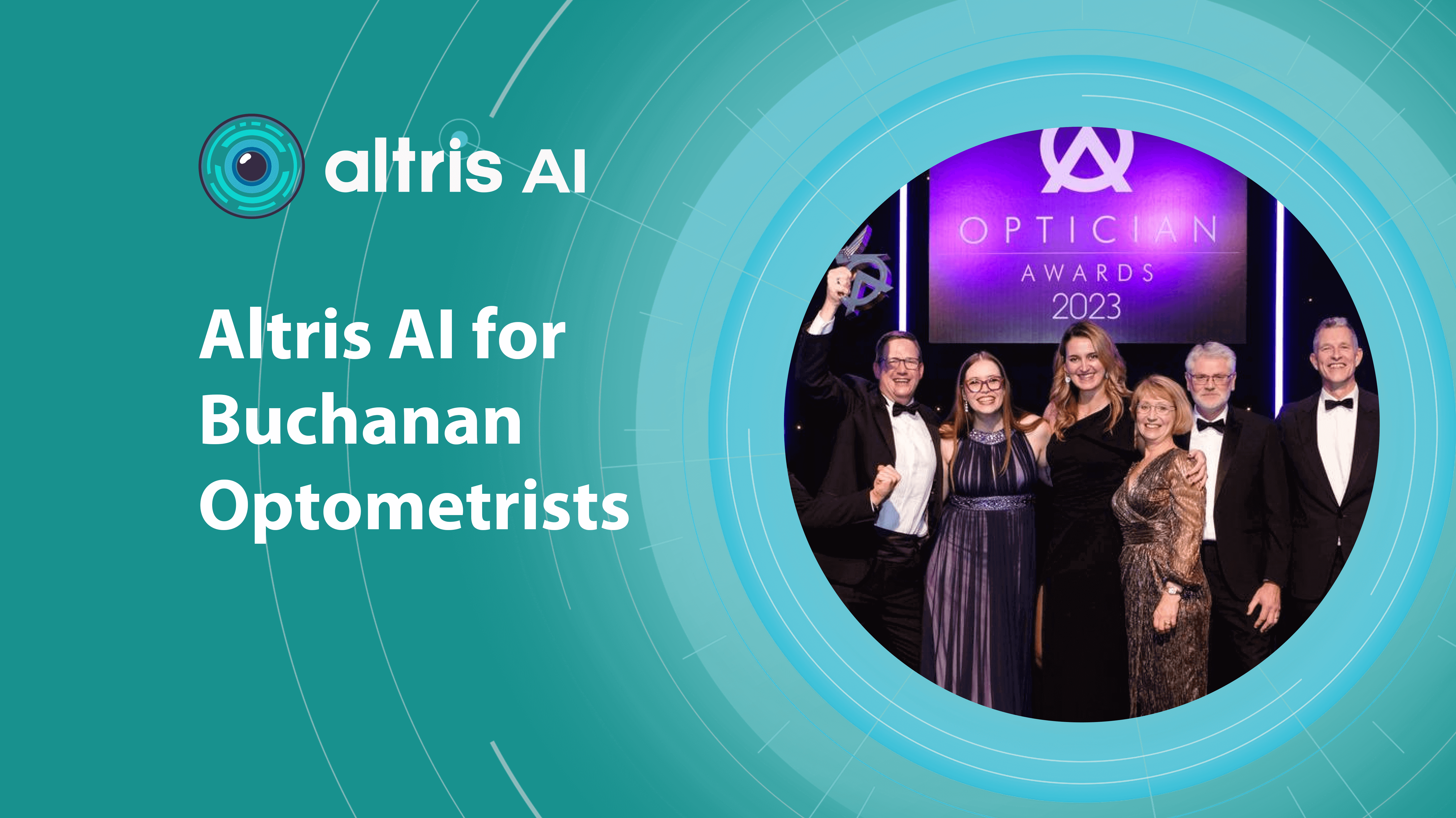


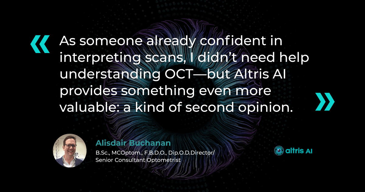
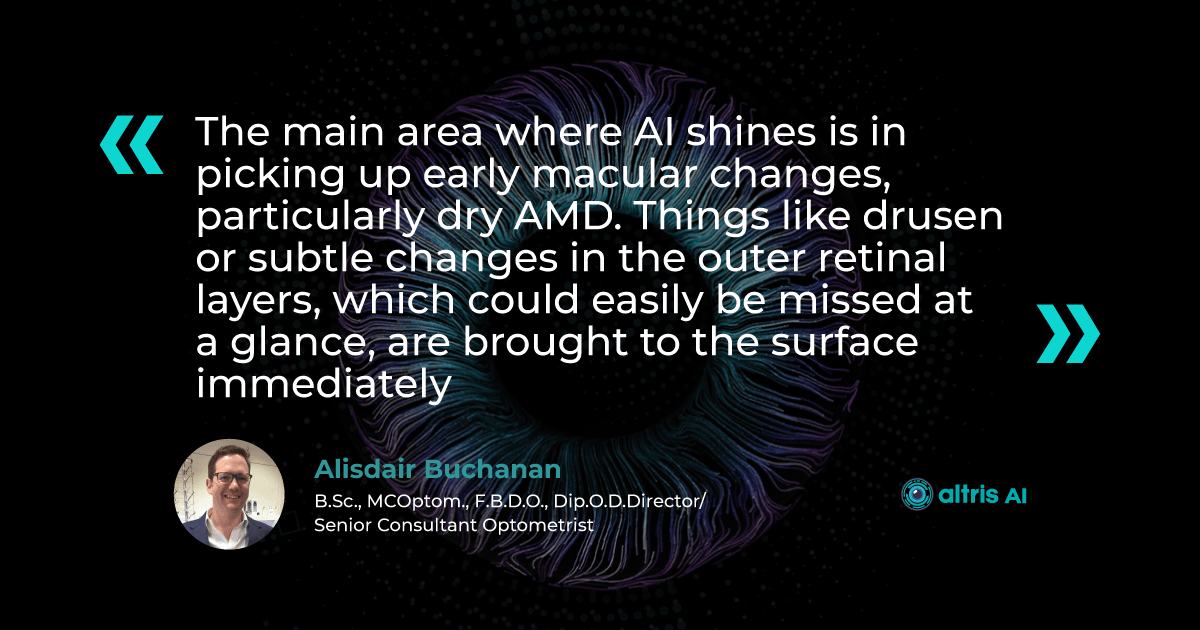
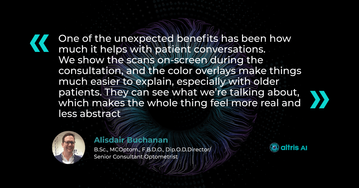
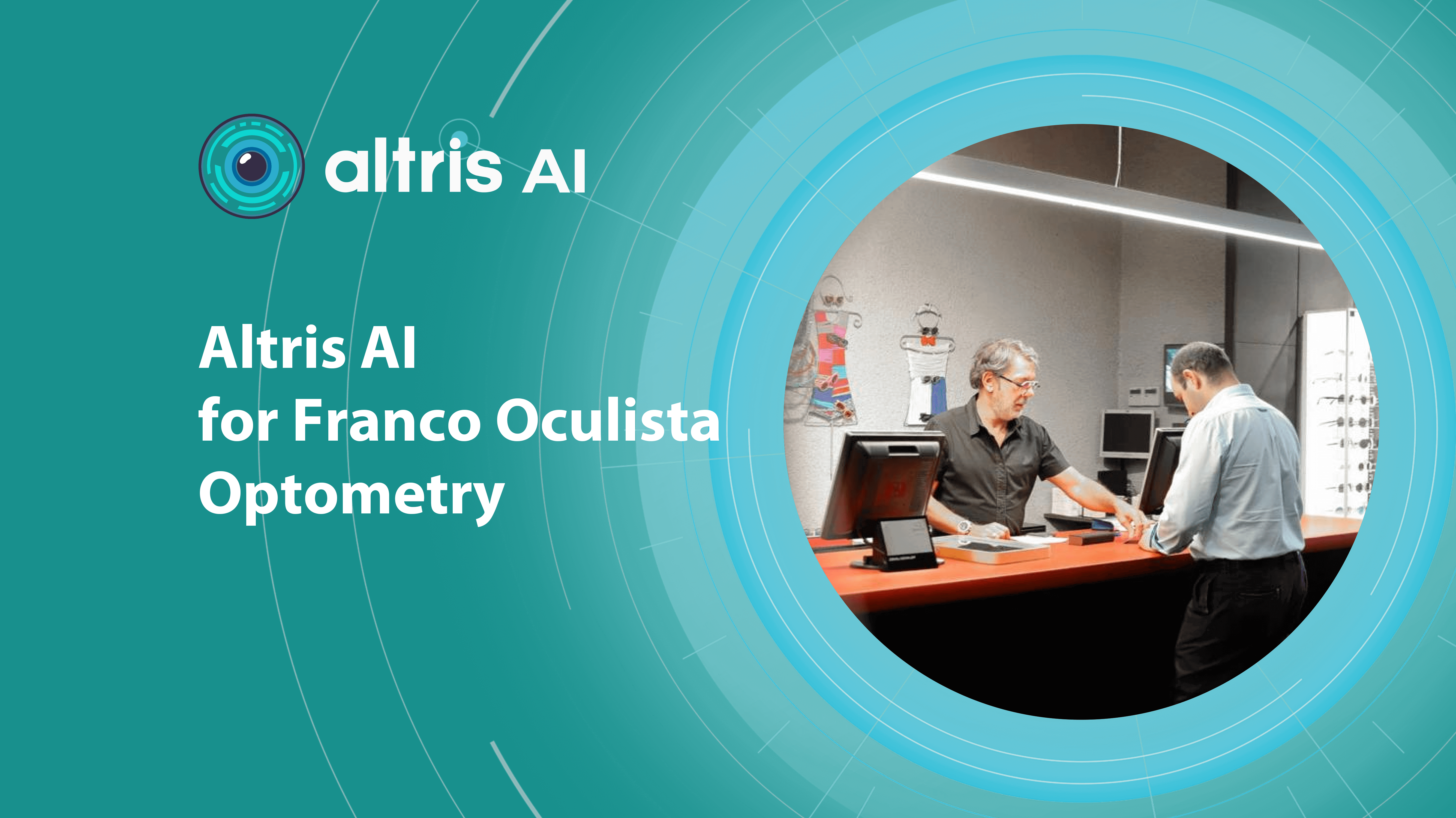


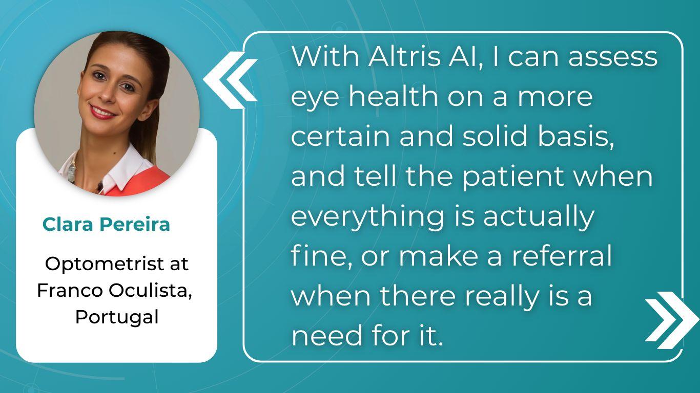
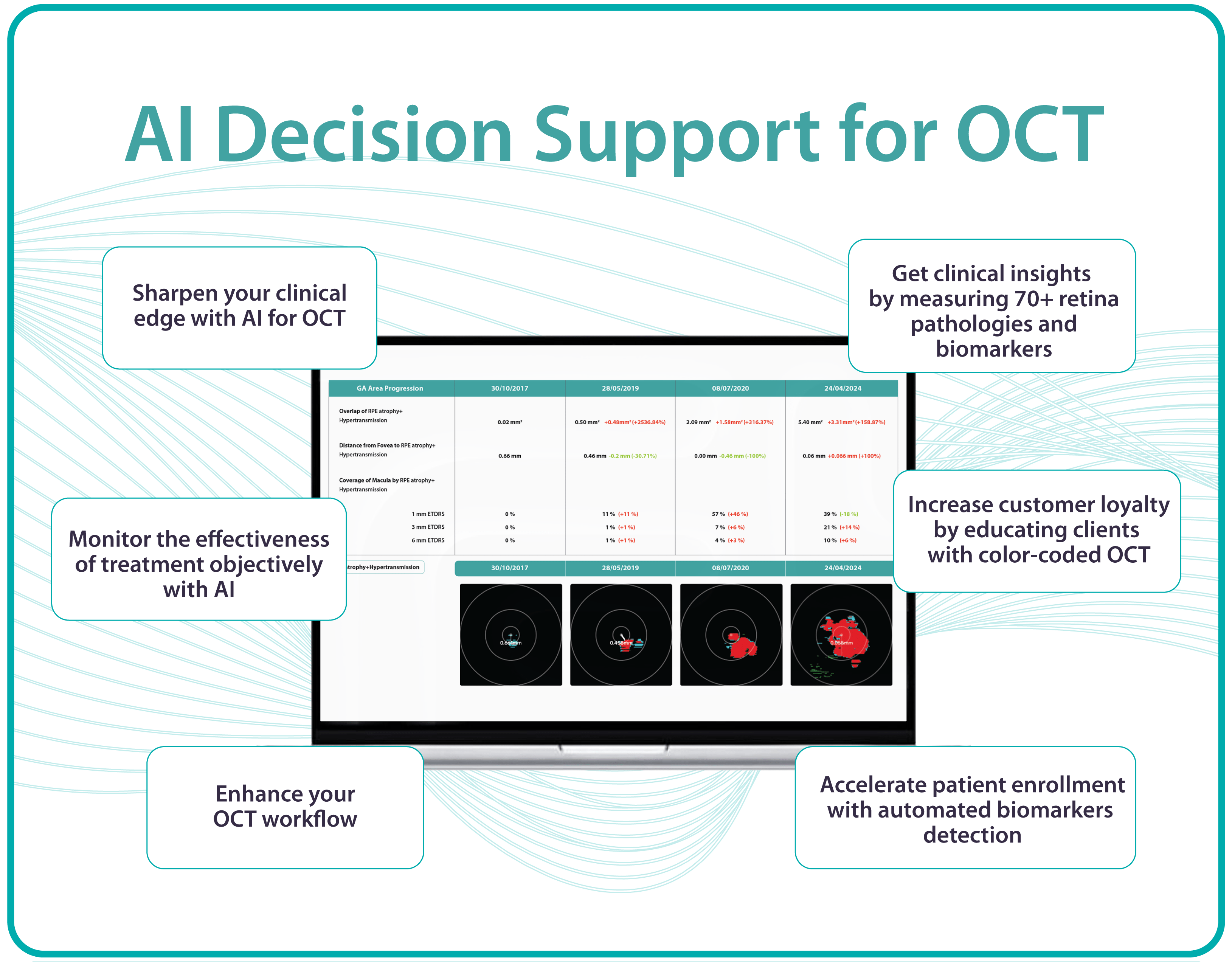
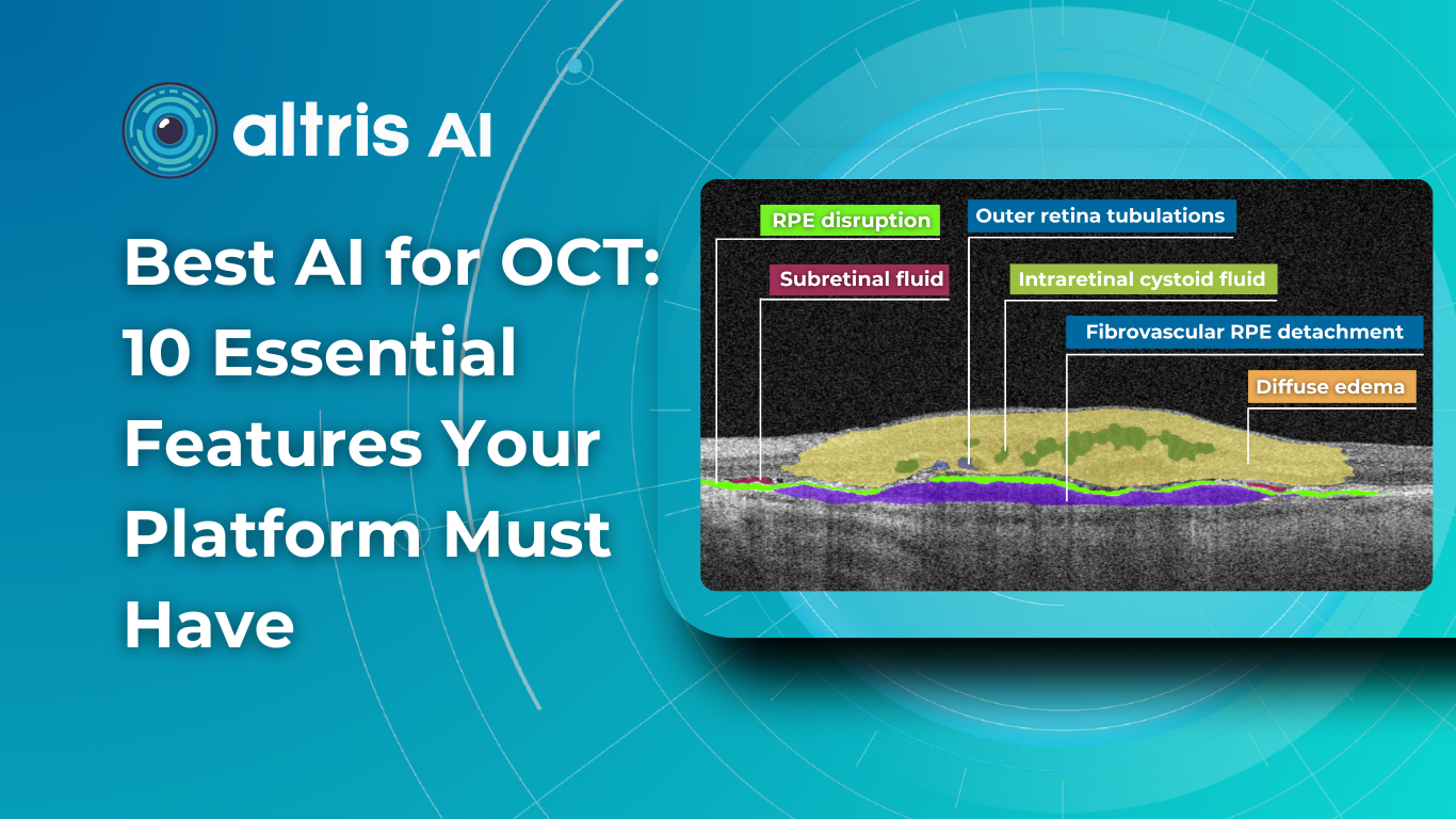
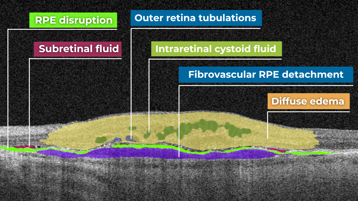

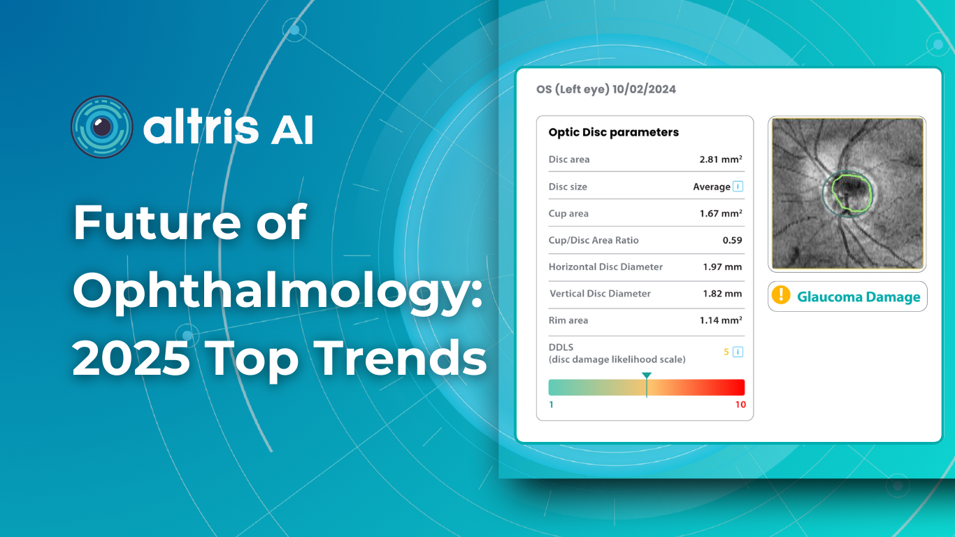
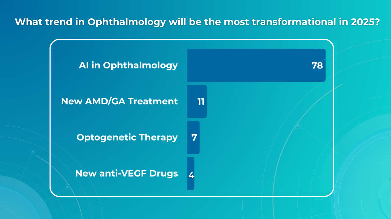
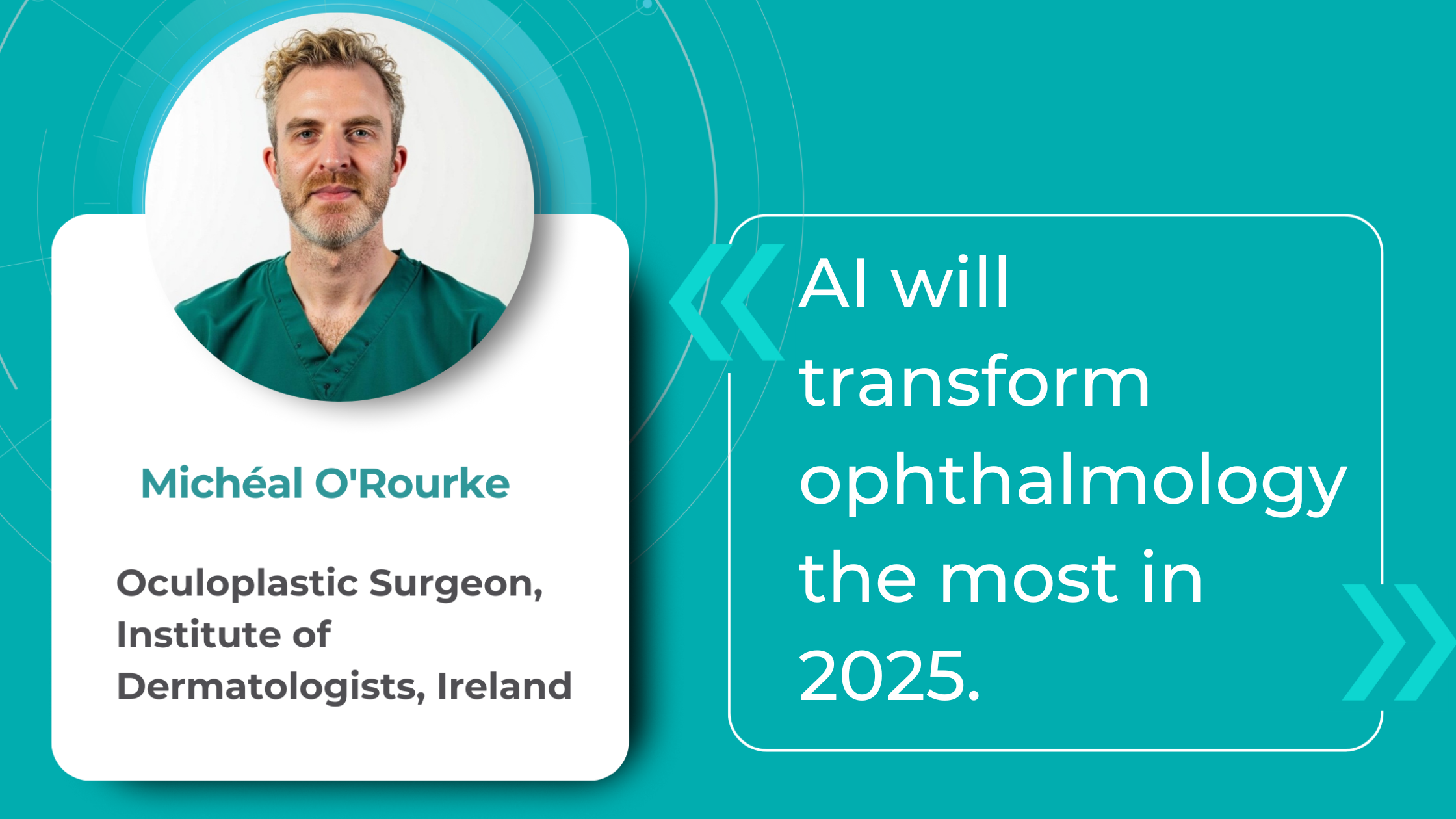
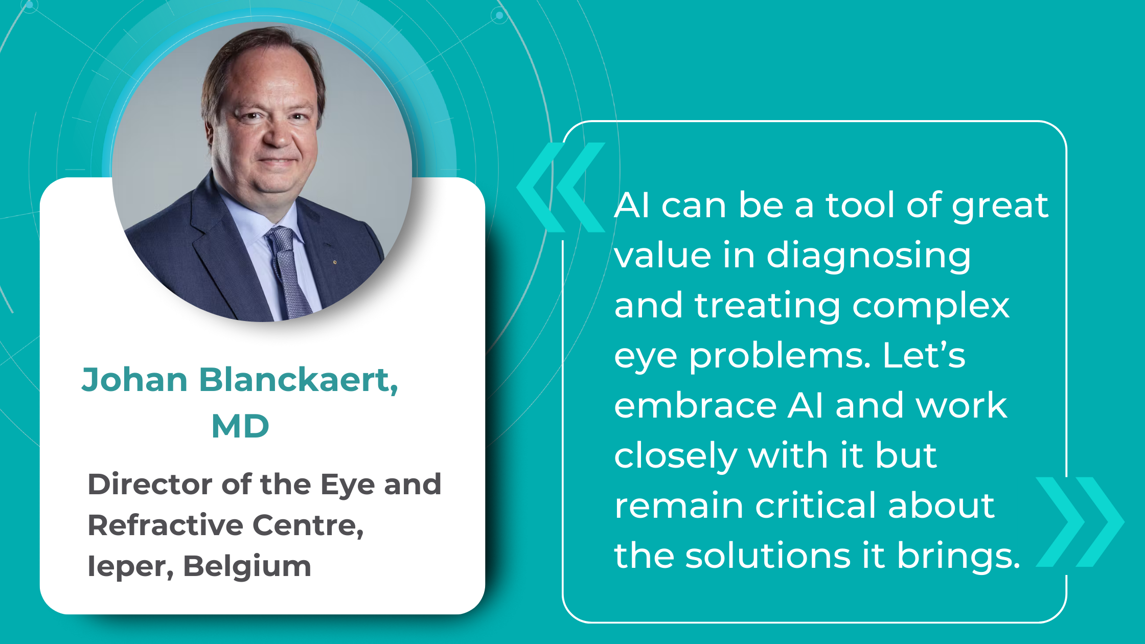
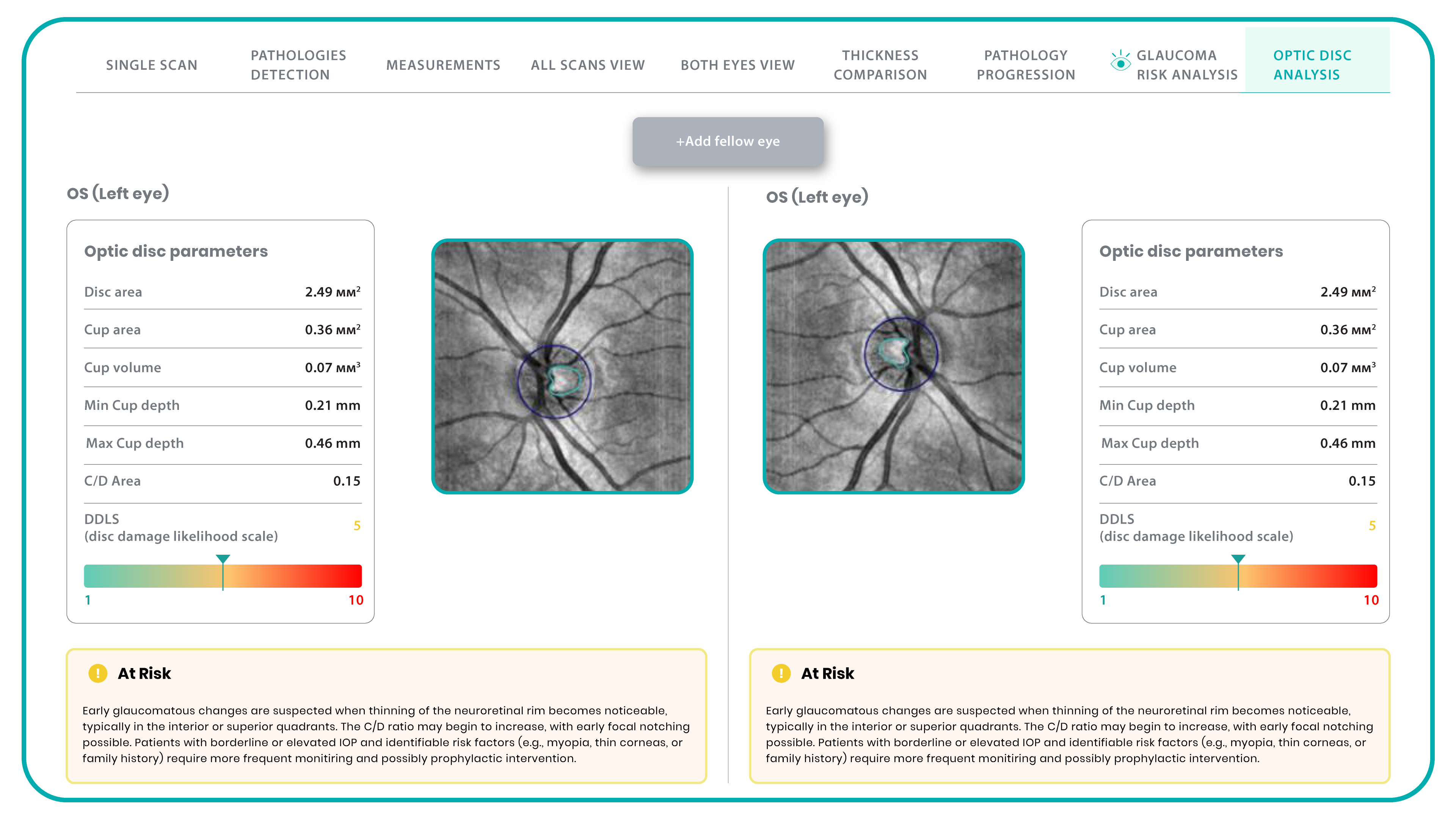
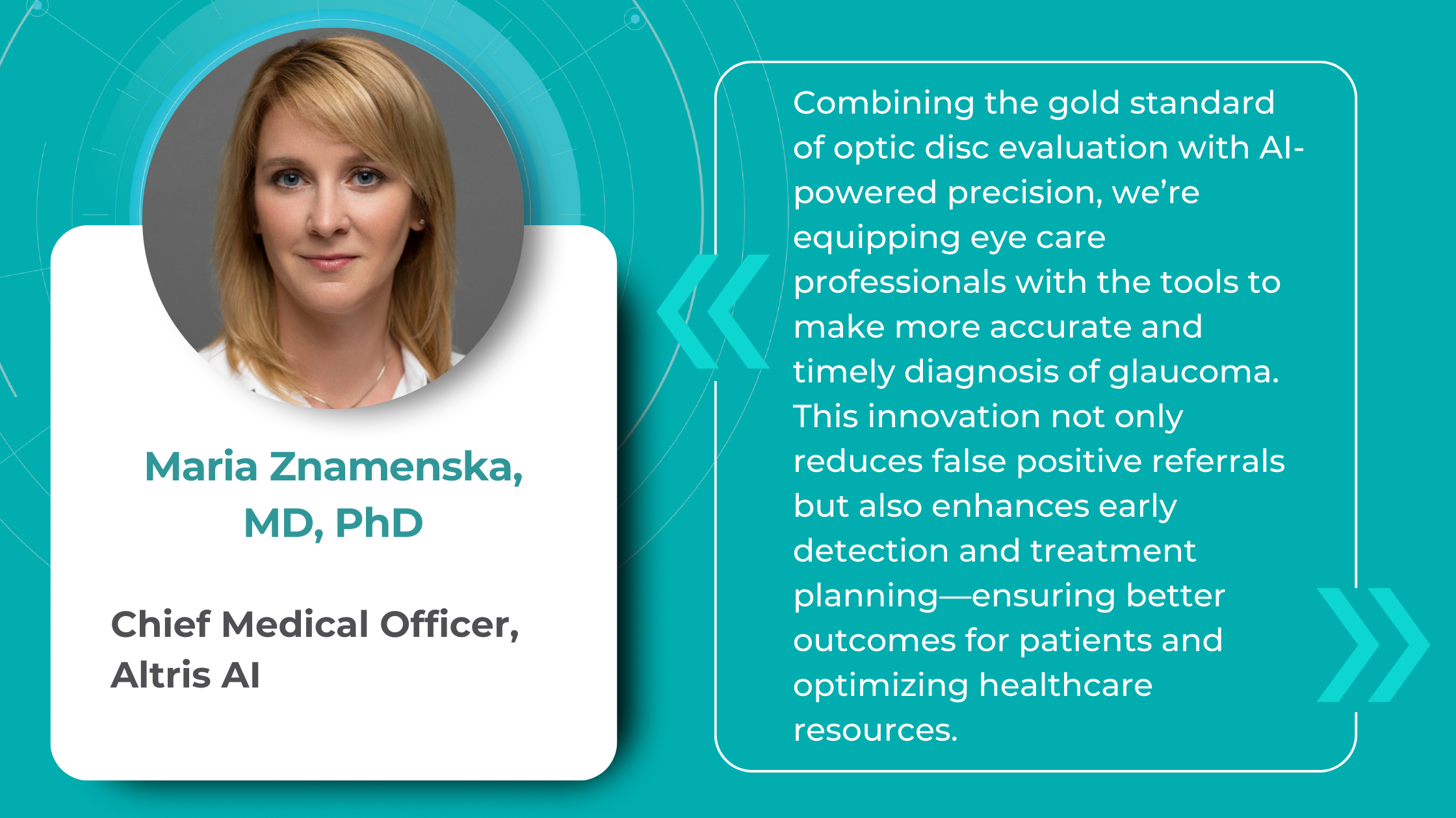
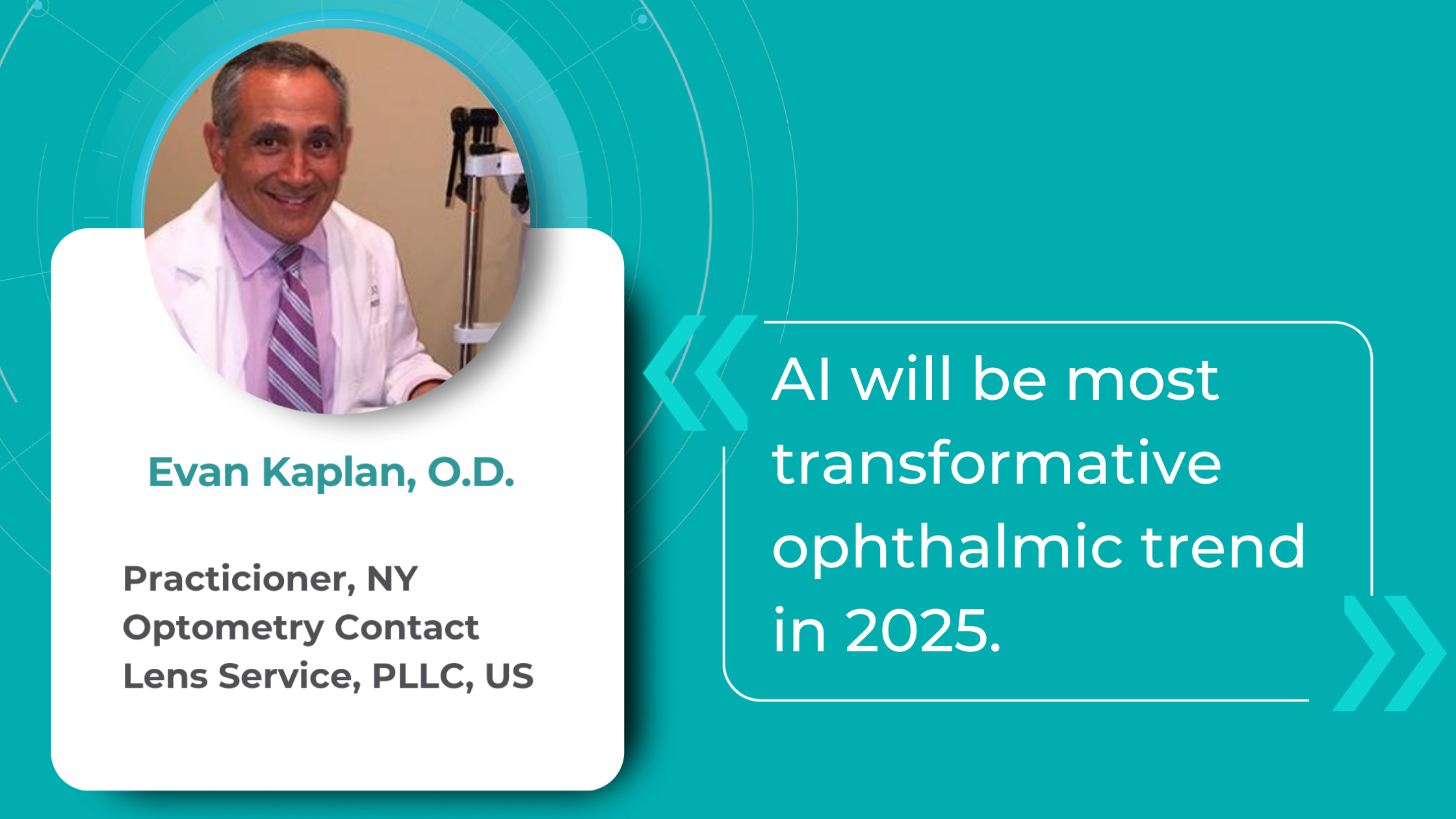
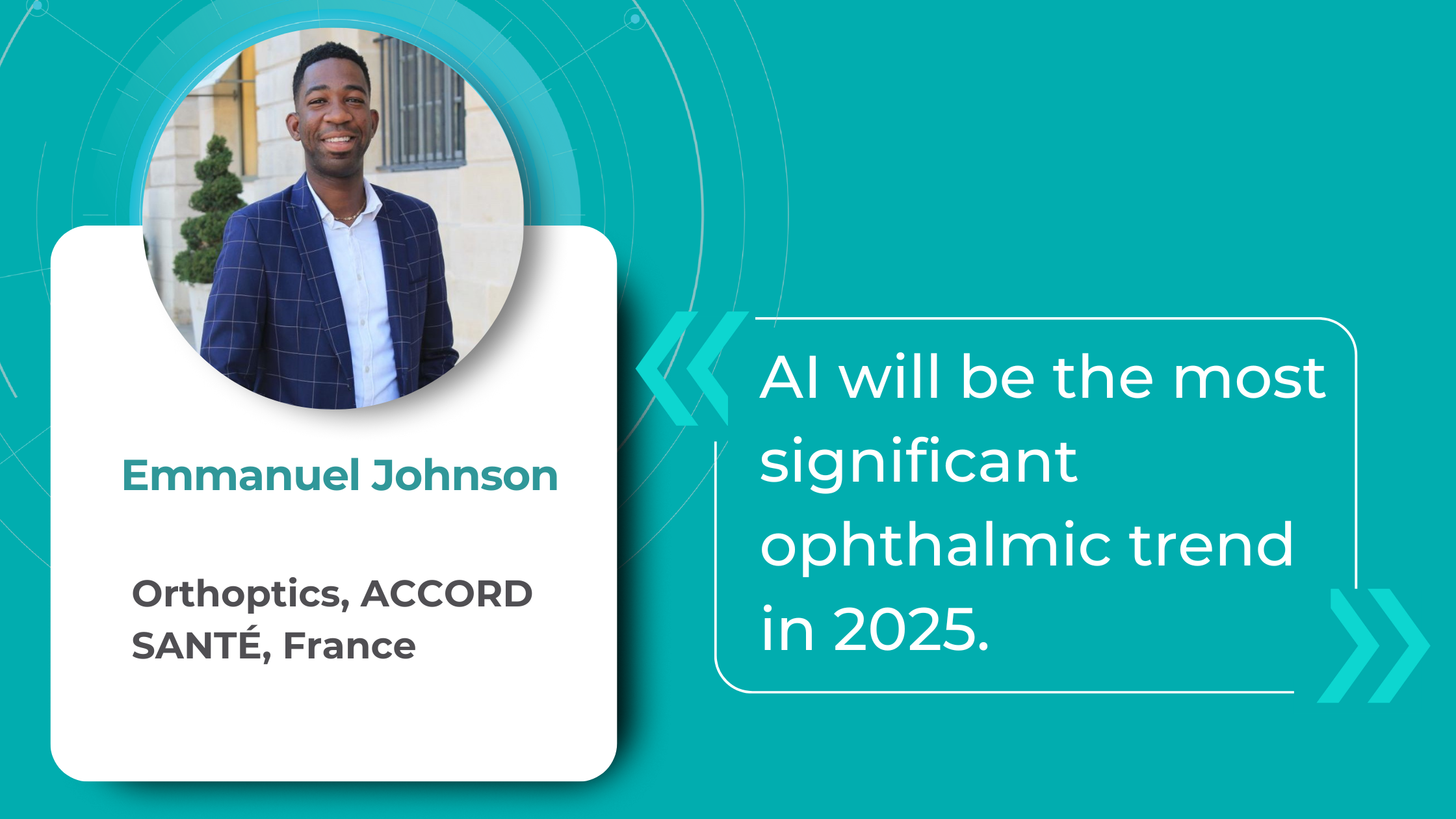
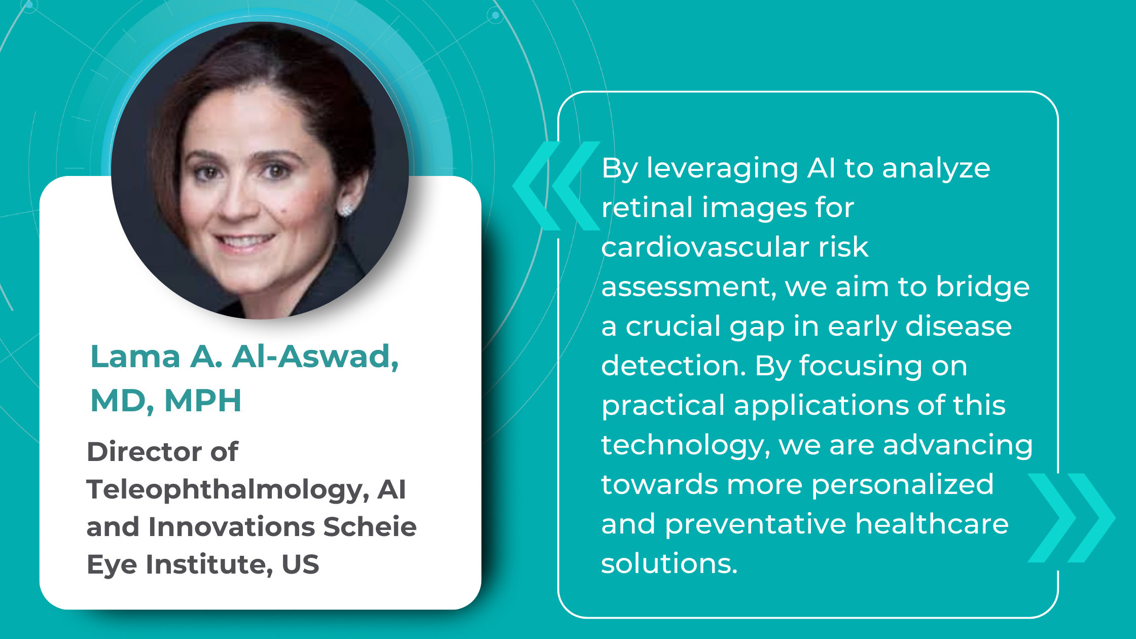
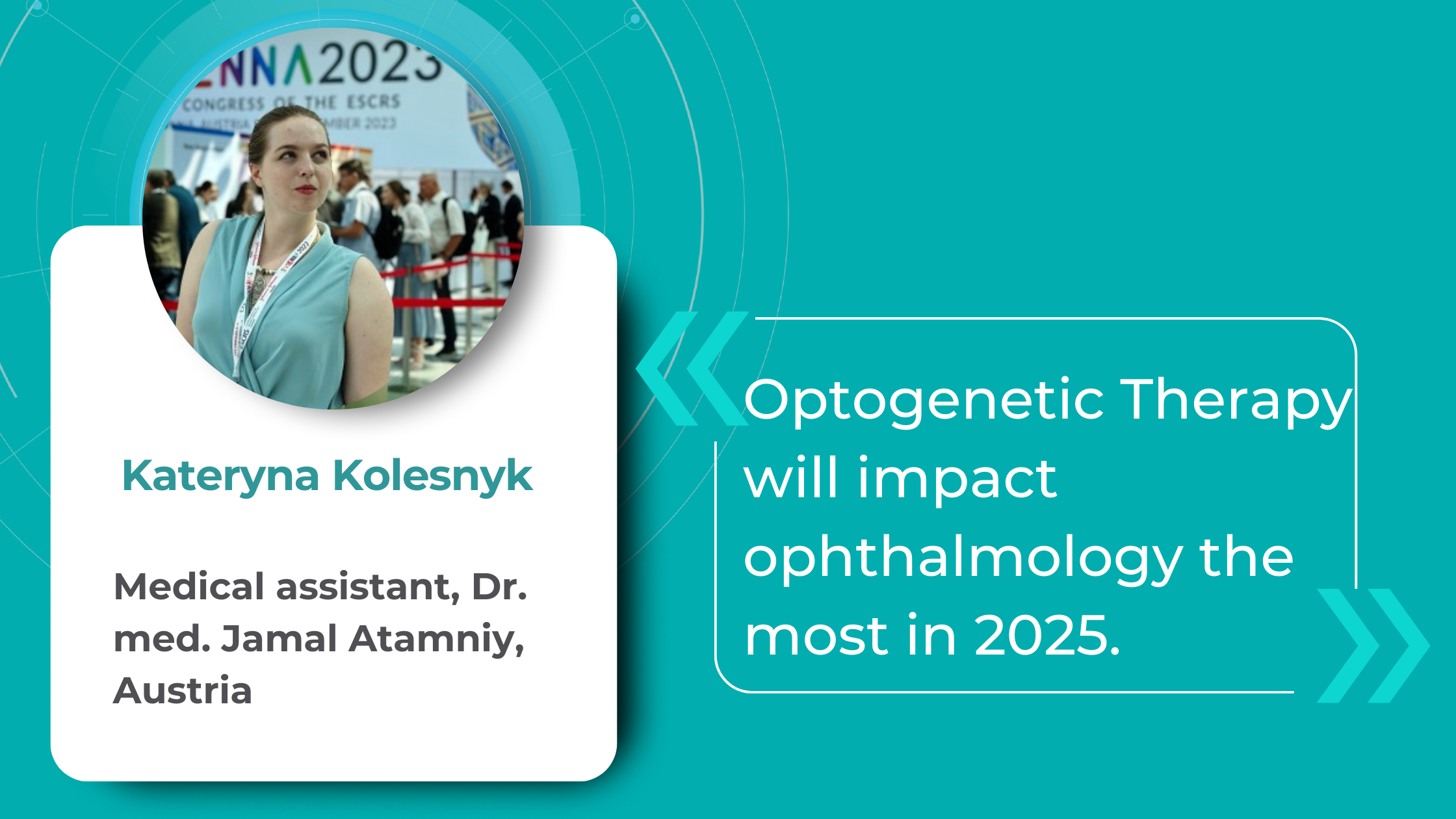
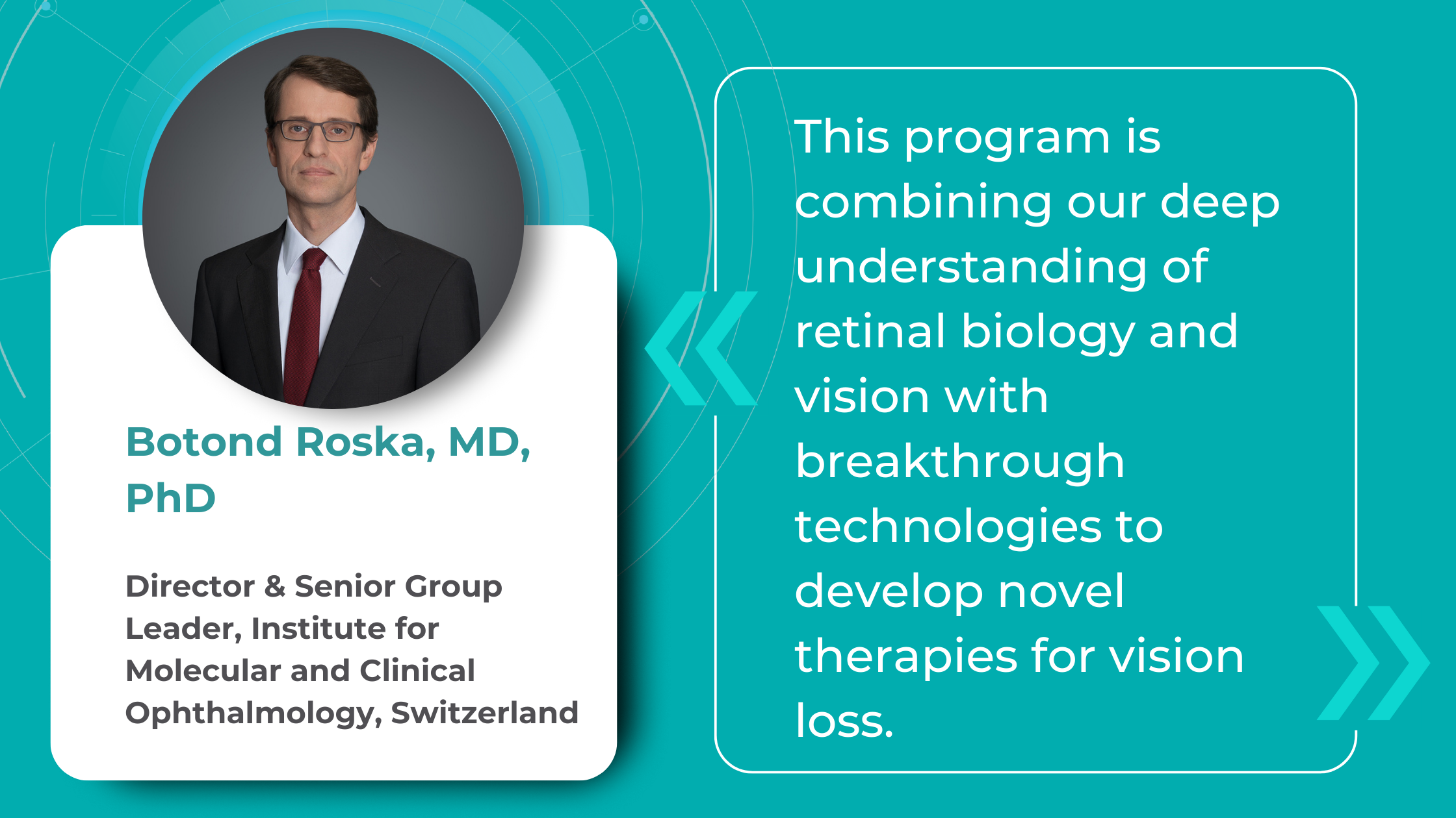
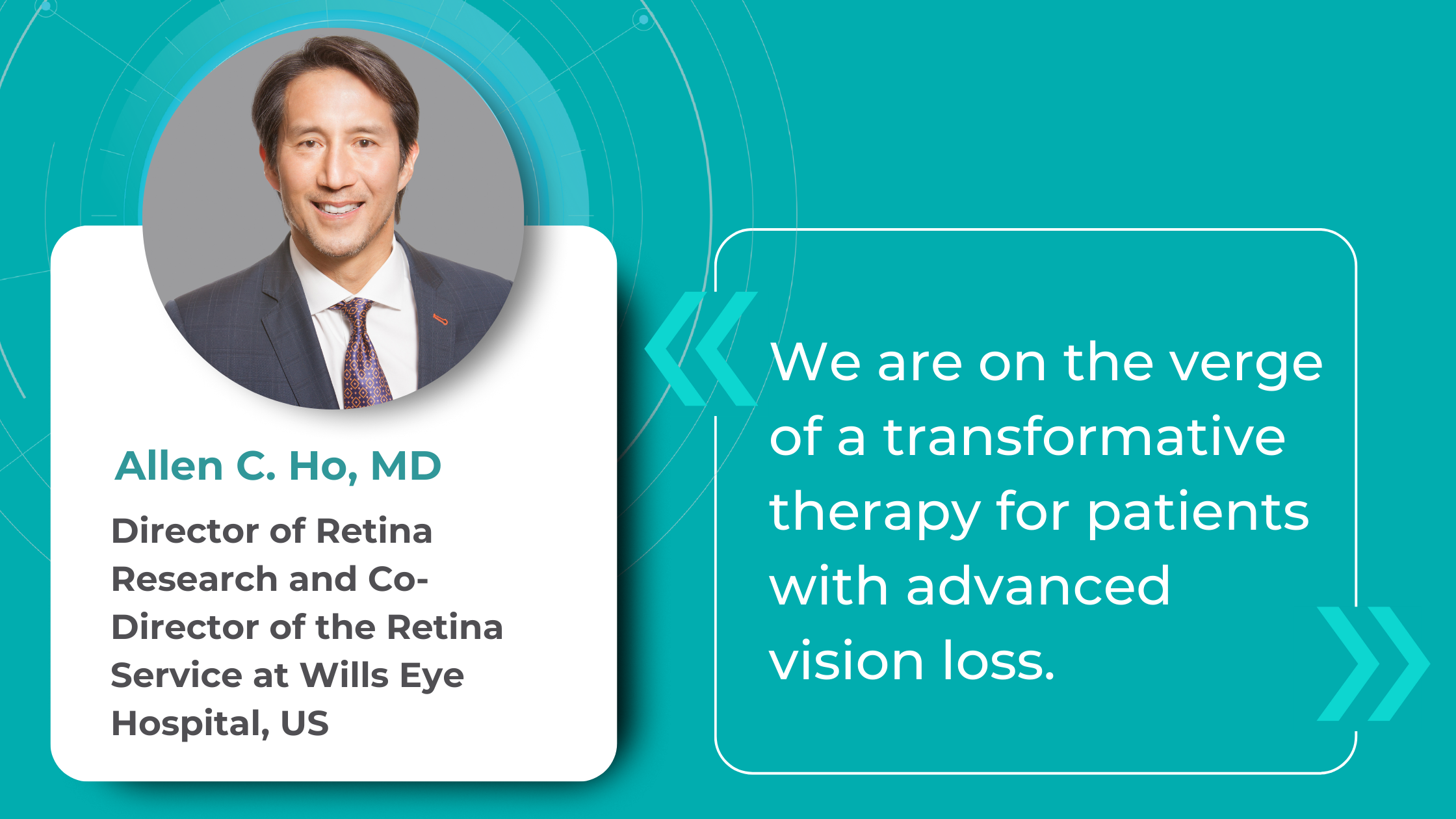
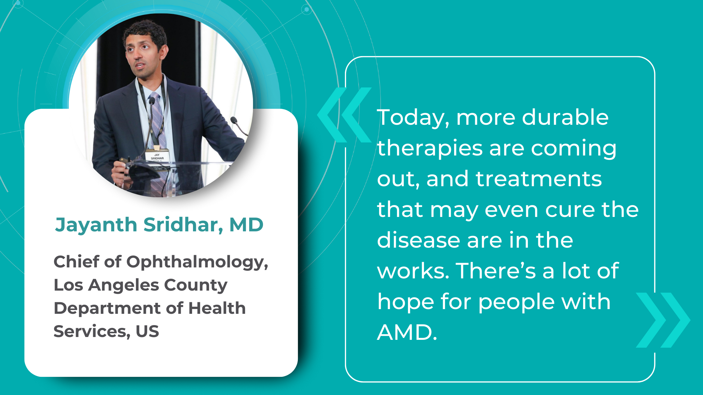
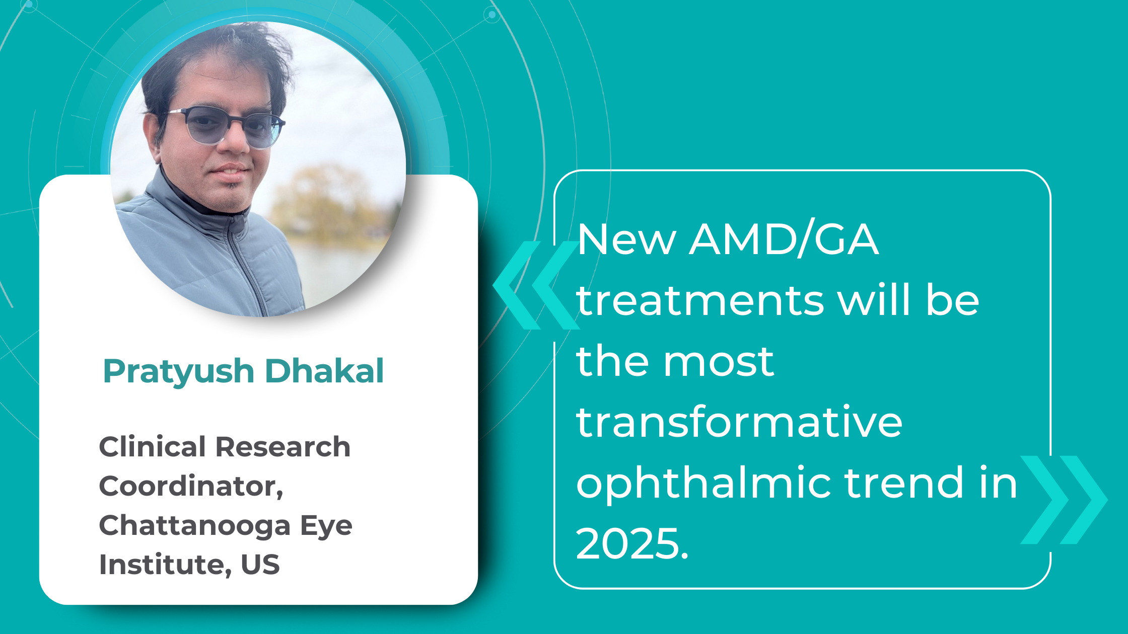
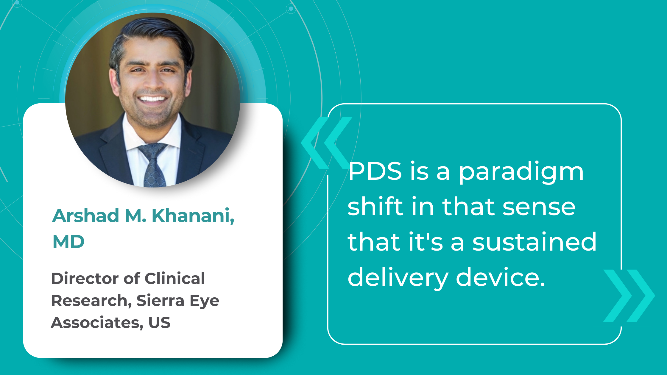
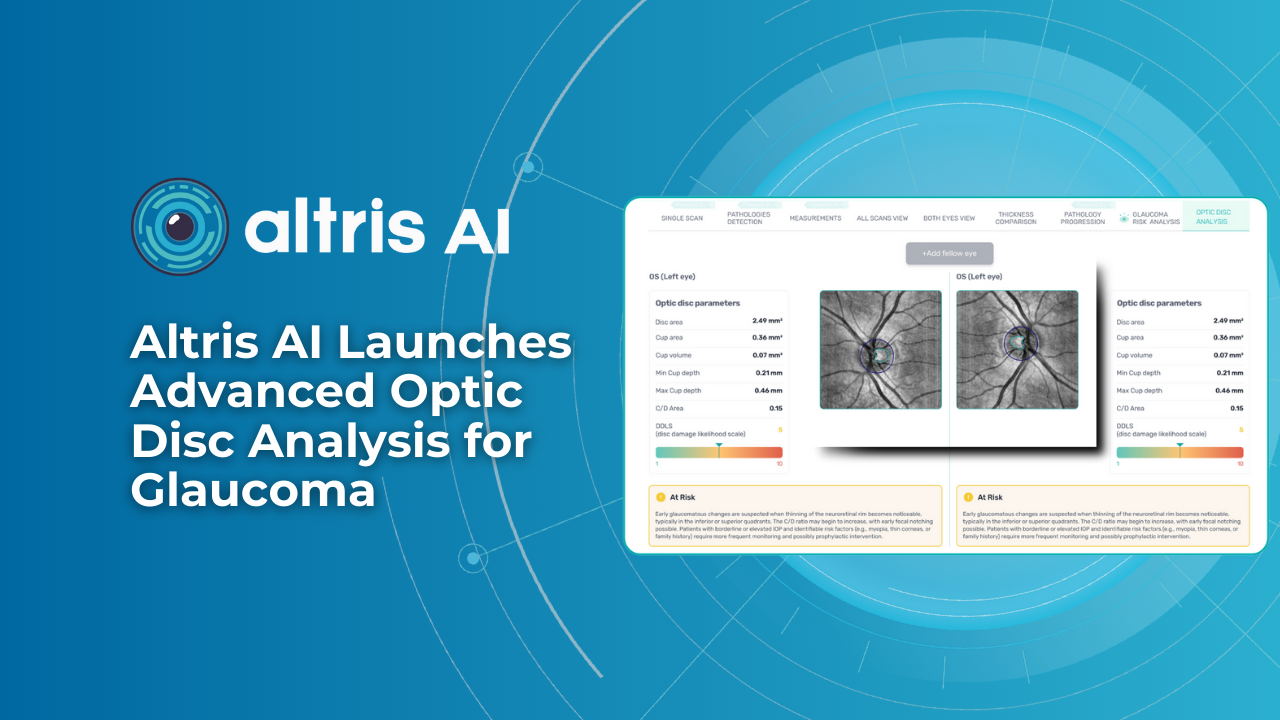
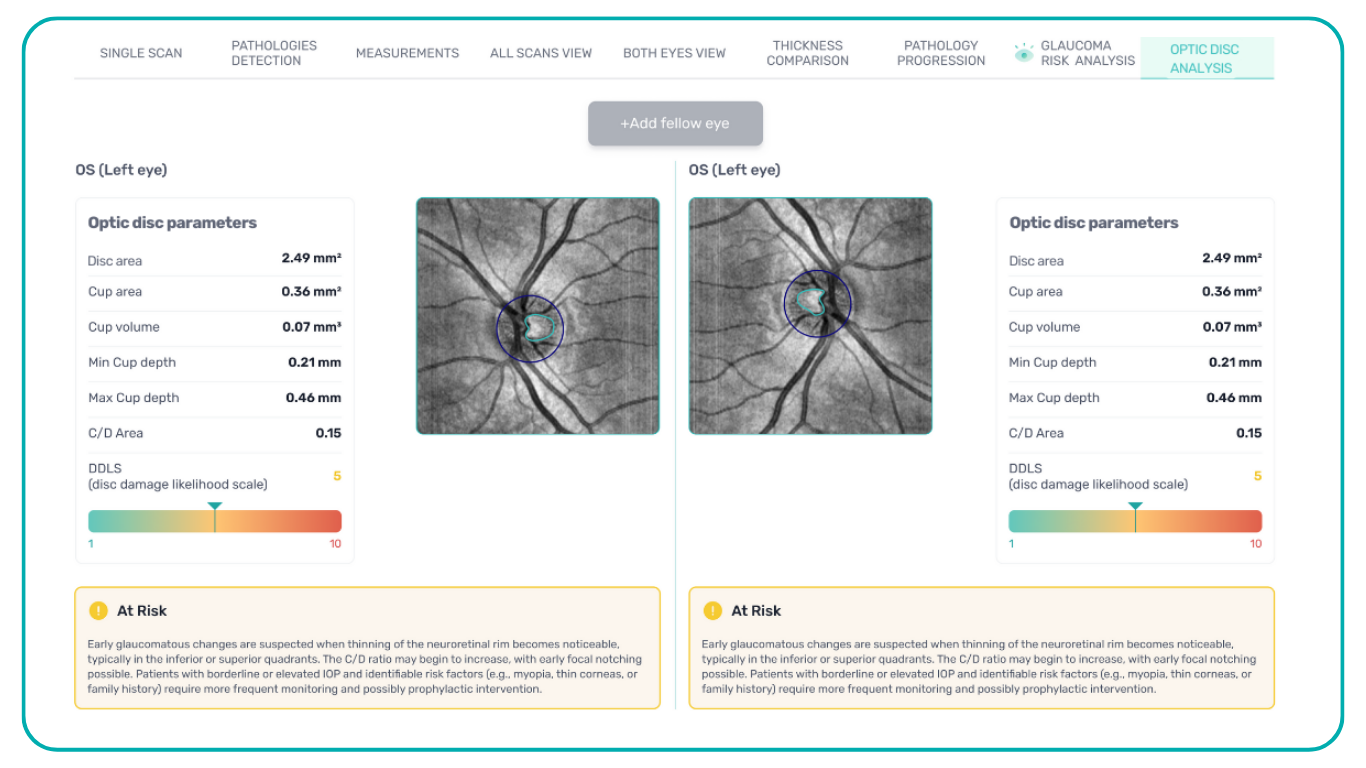
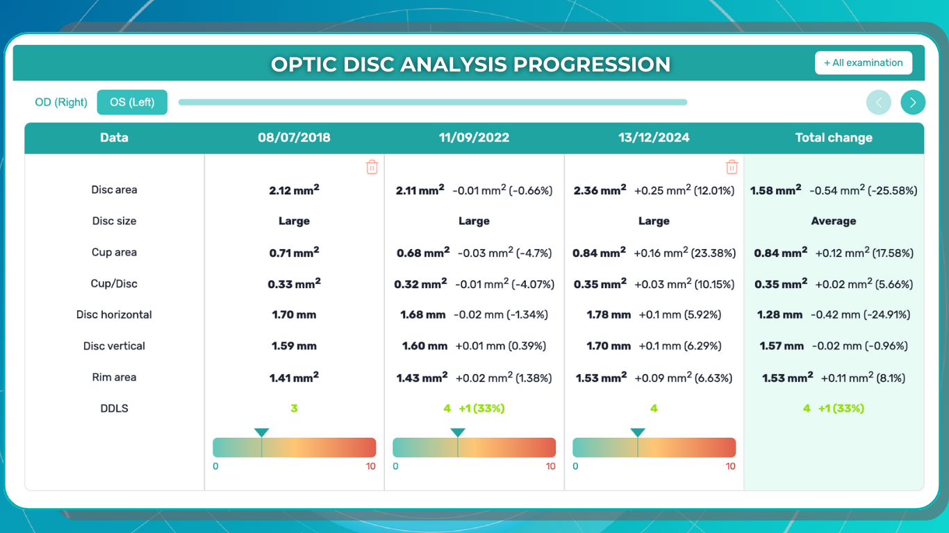

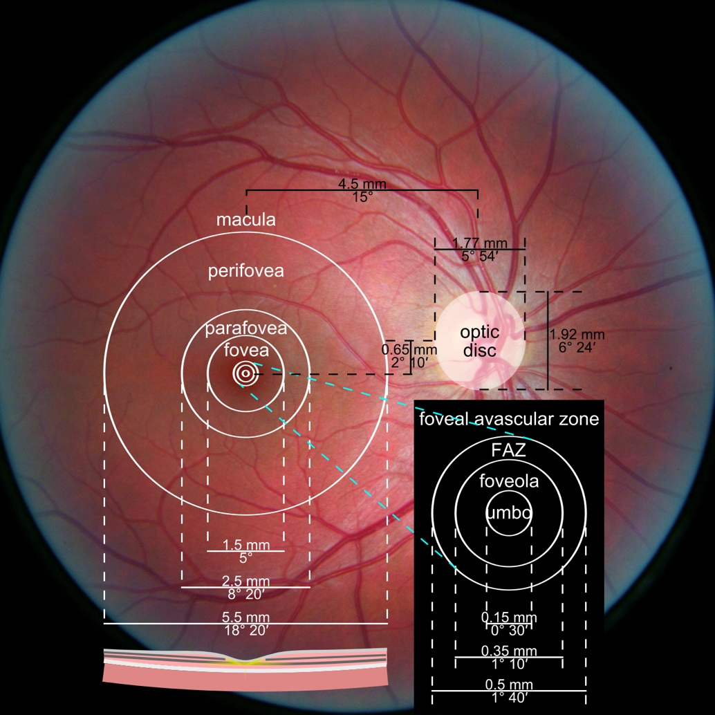
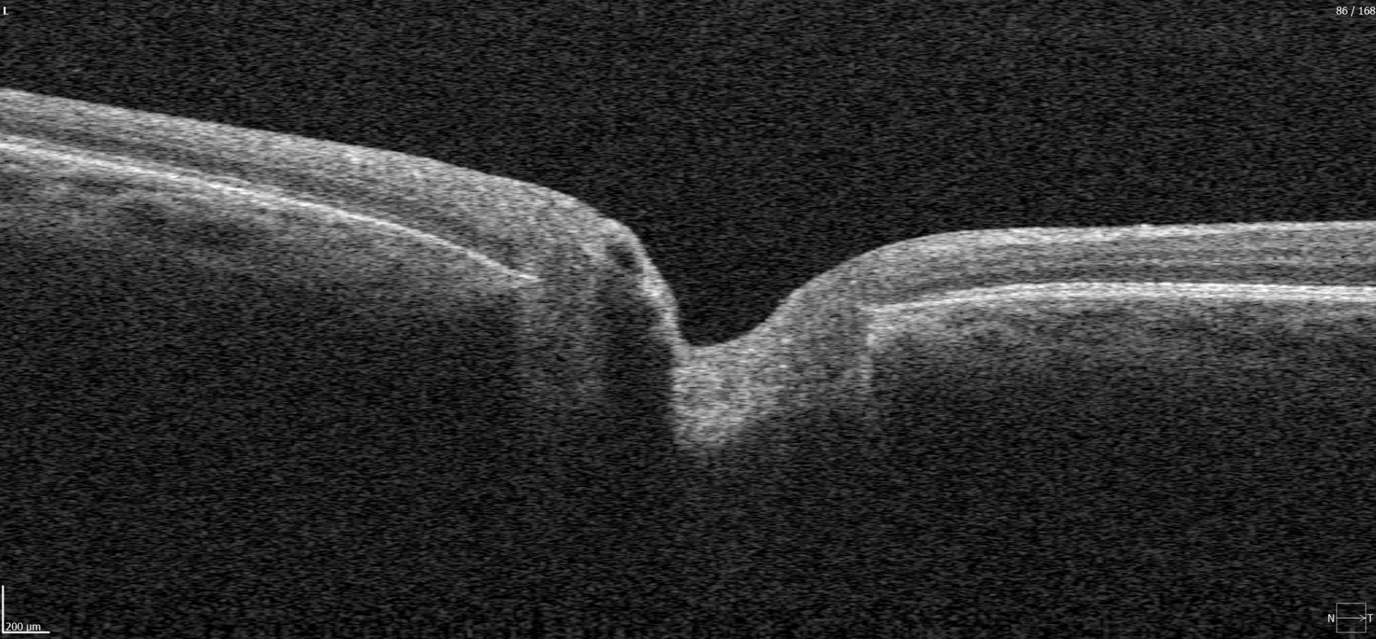
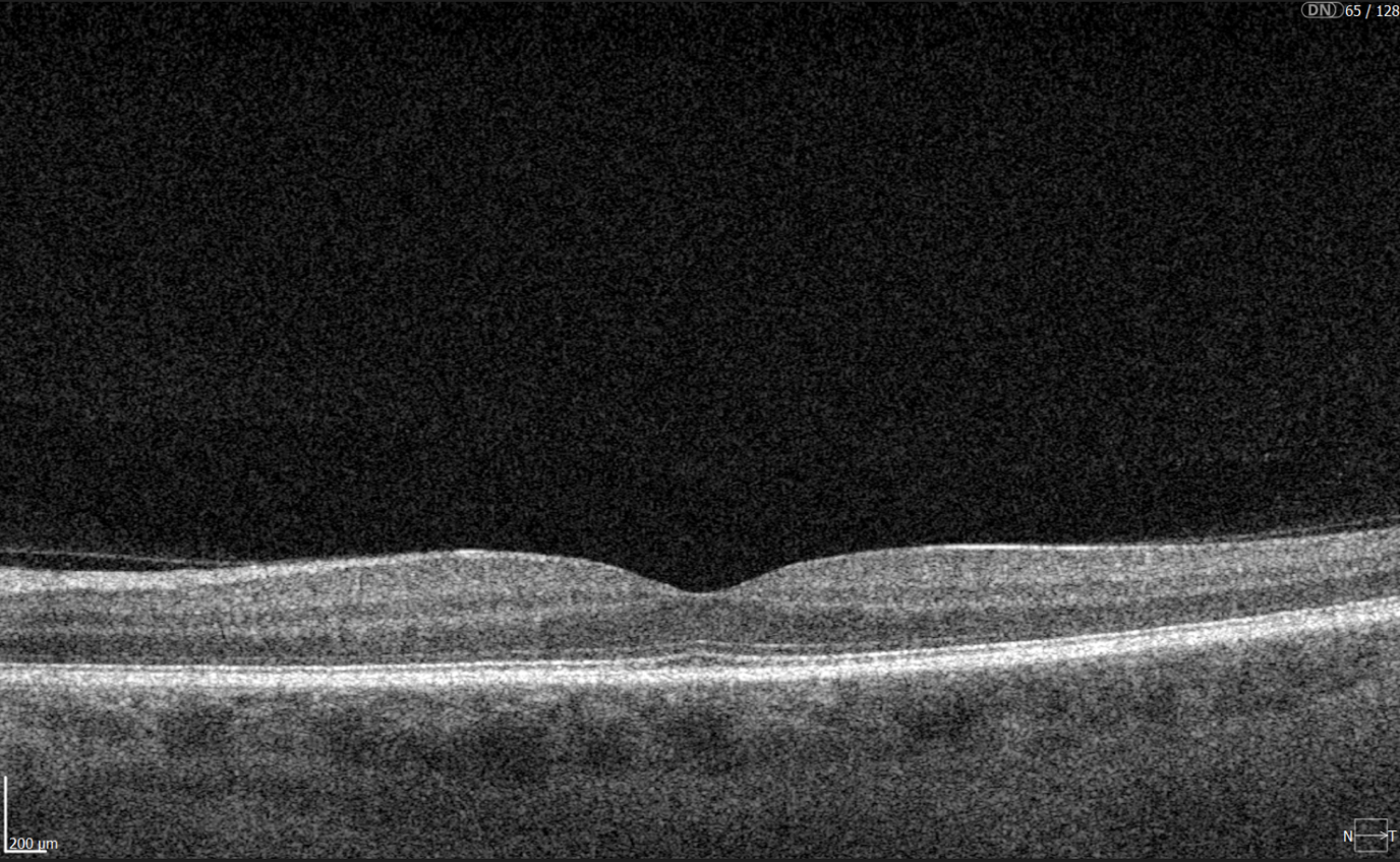
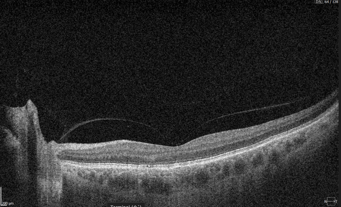
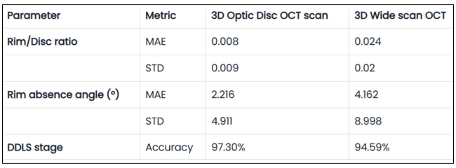
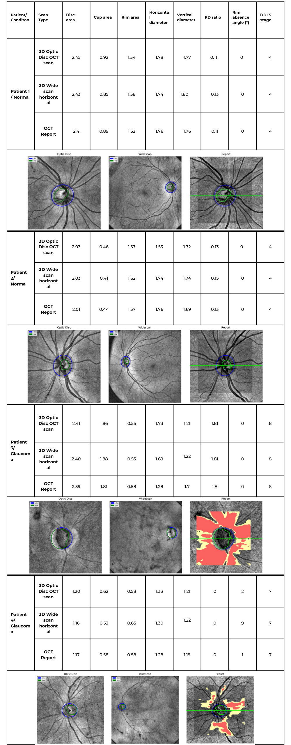
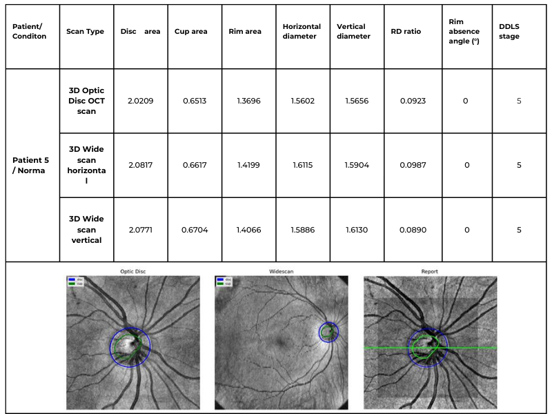
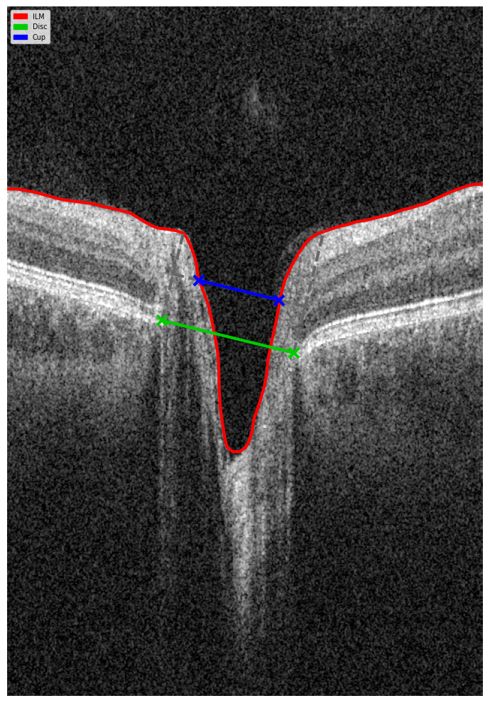
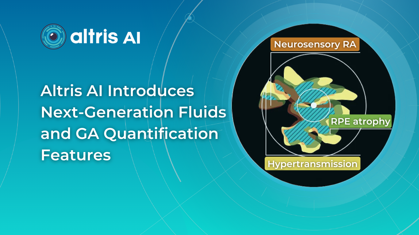
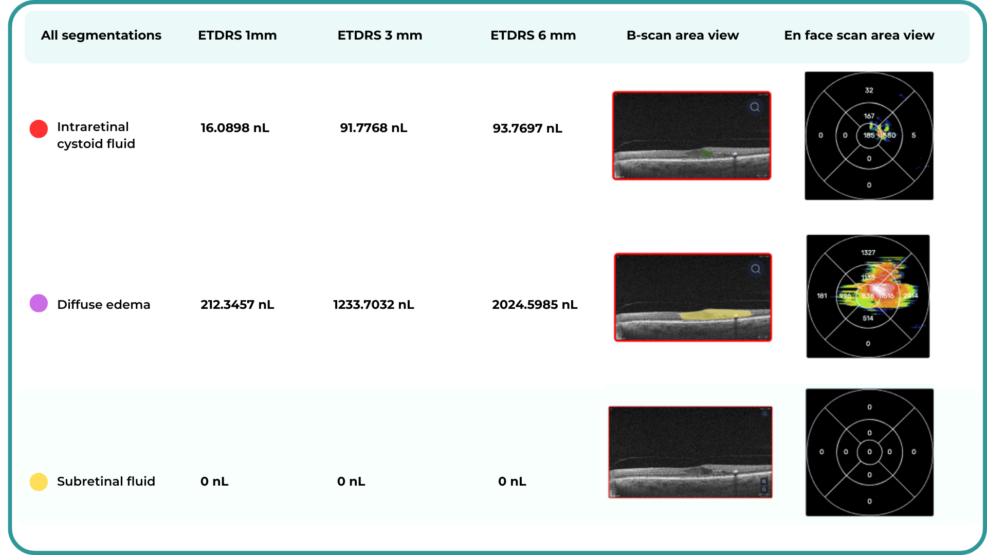
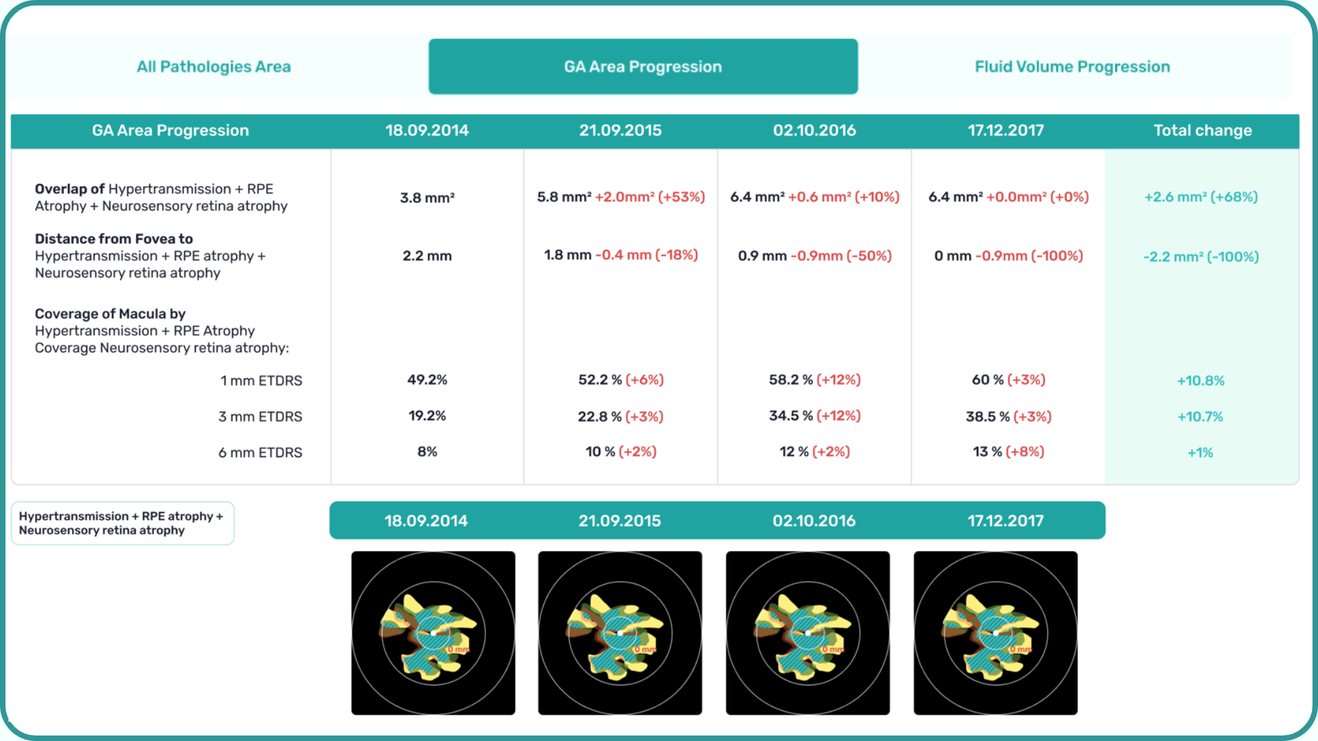
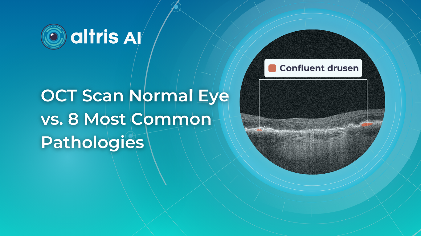
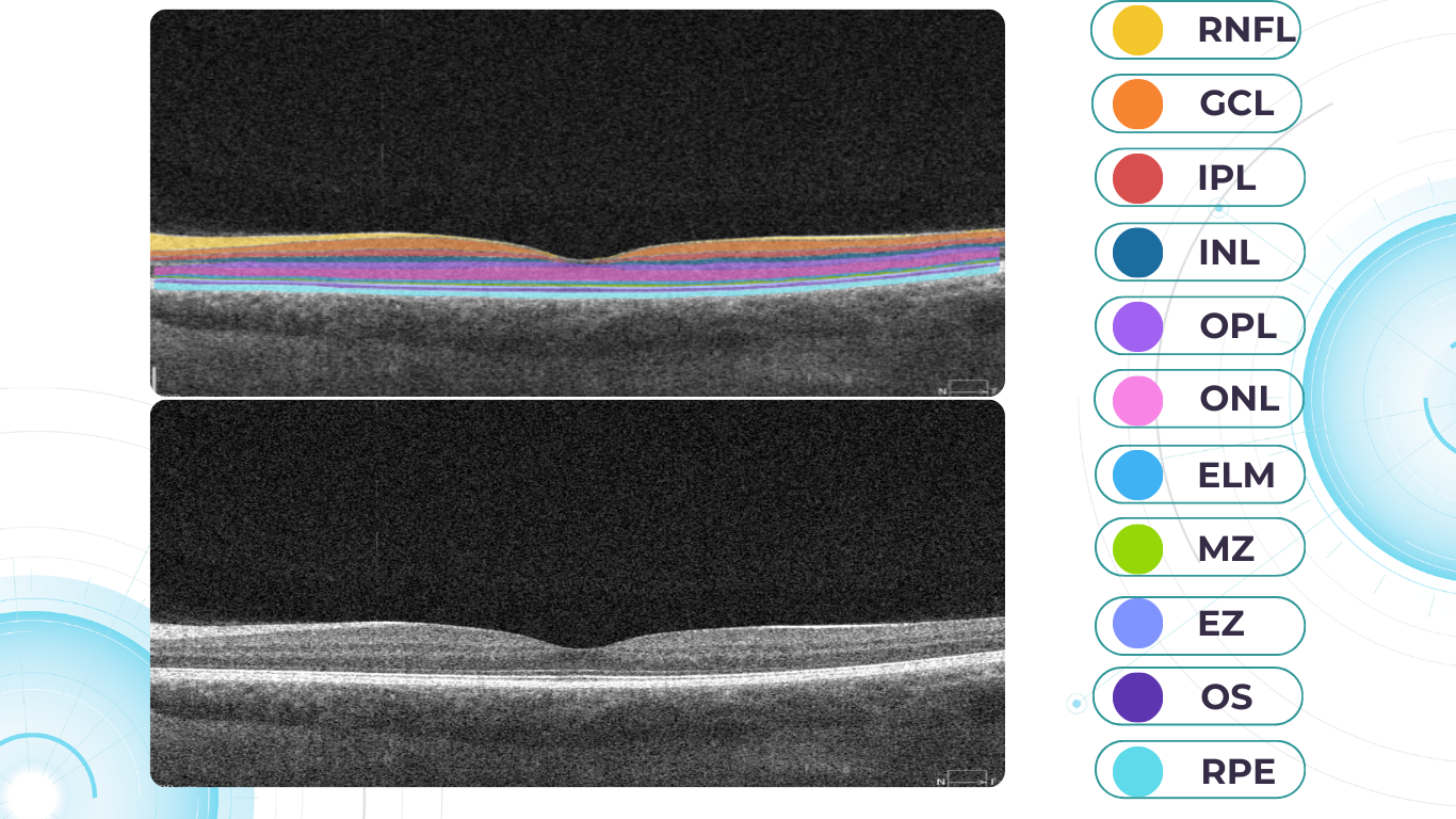
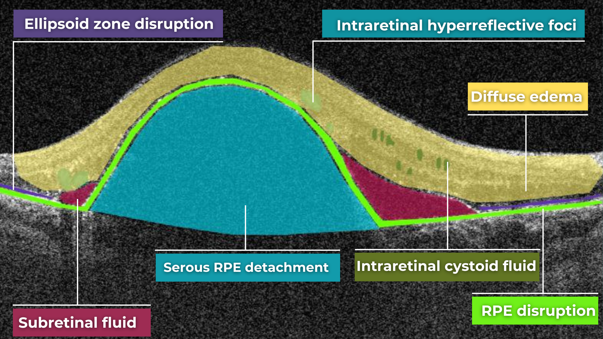
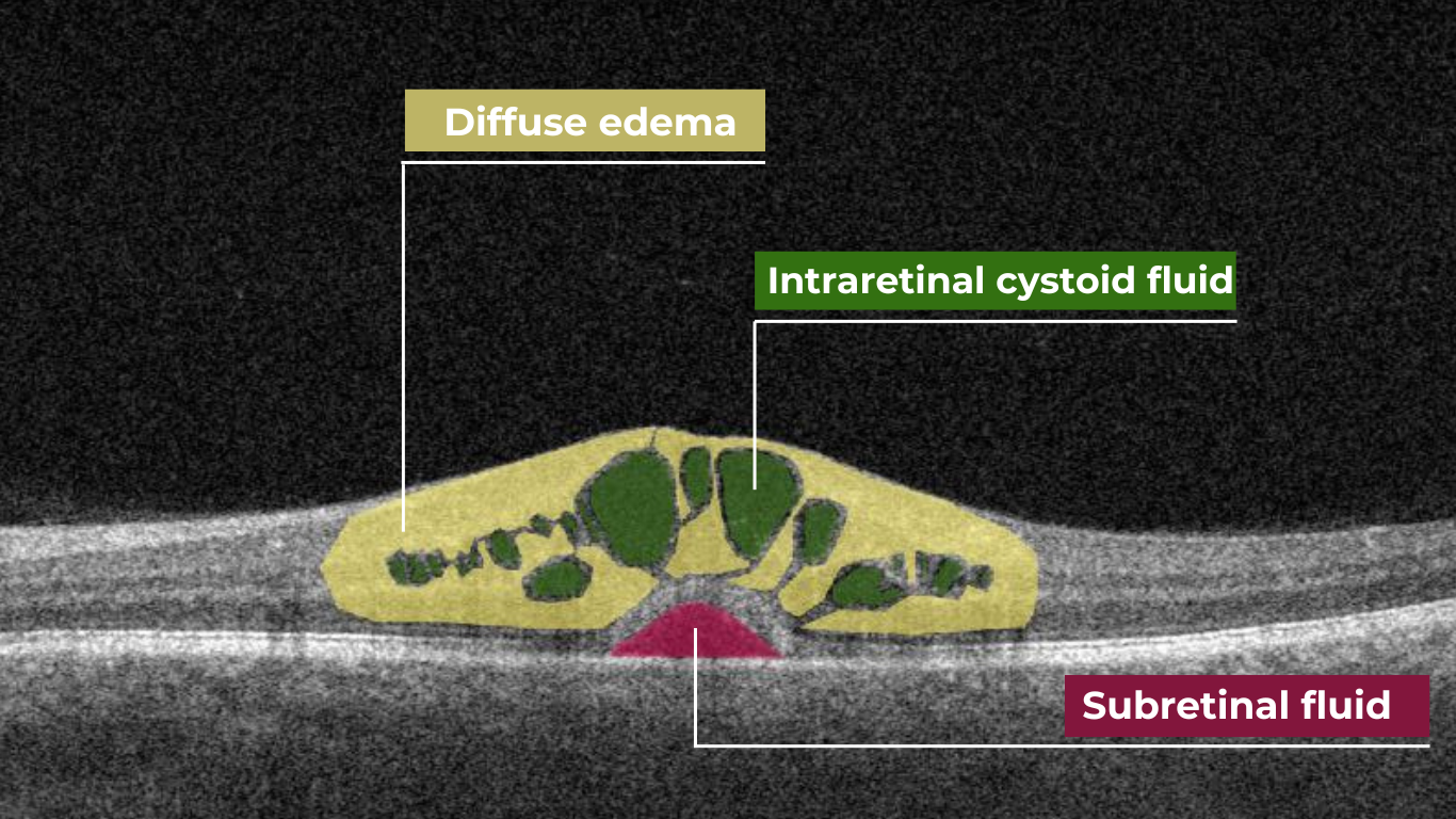

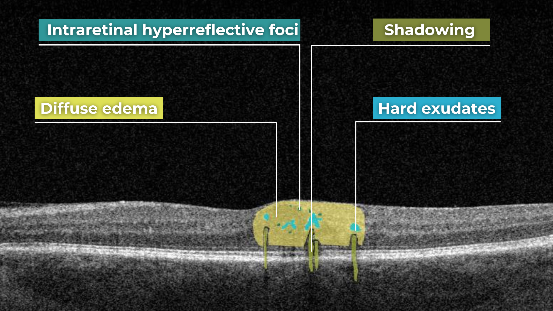
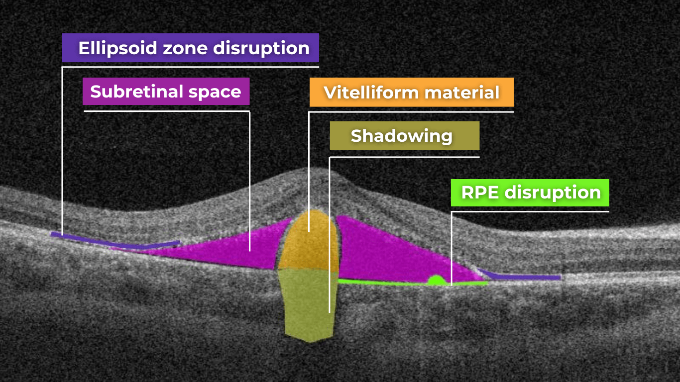


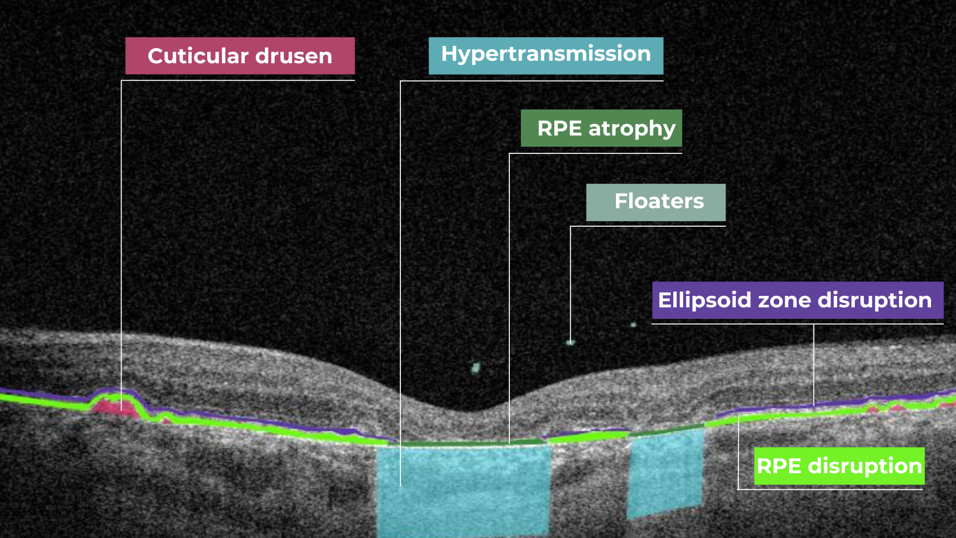 Hypertransmission on OCT
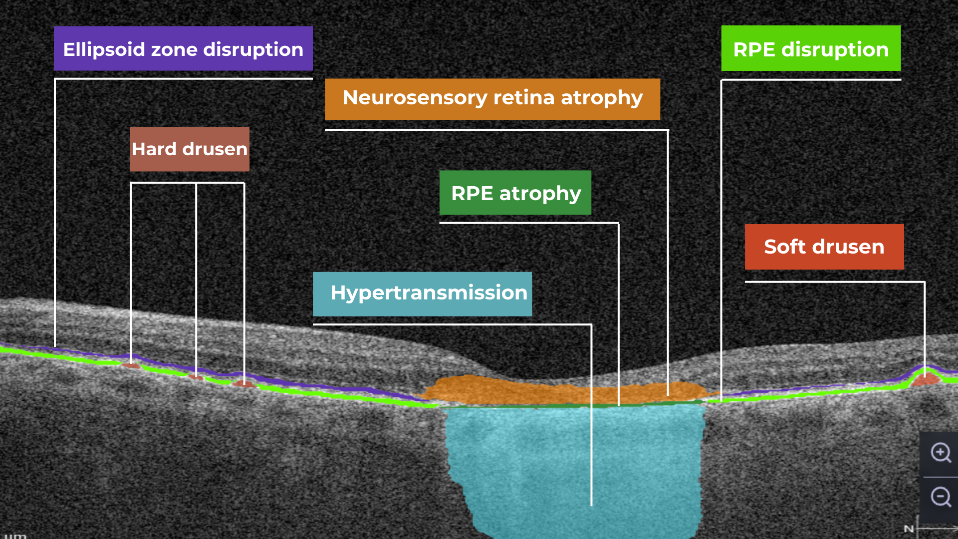
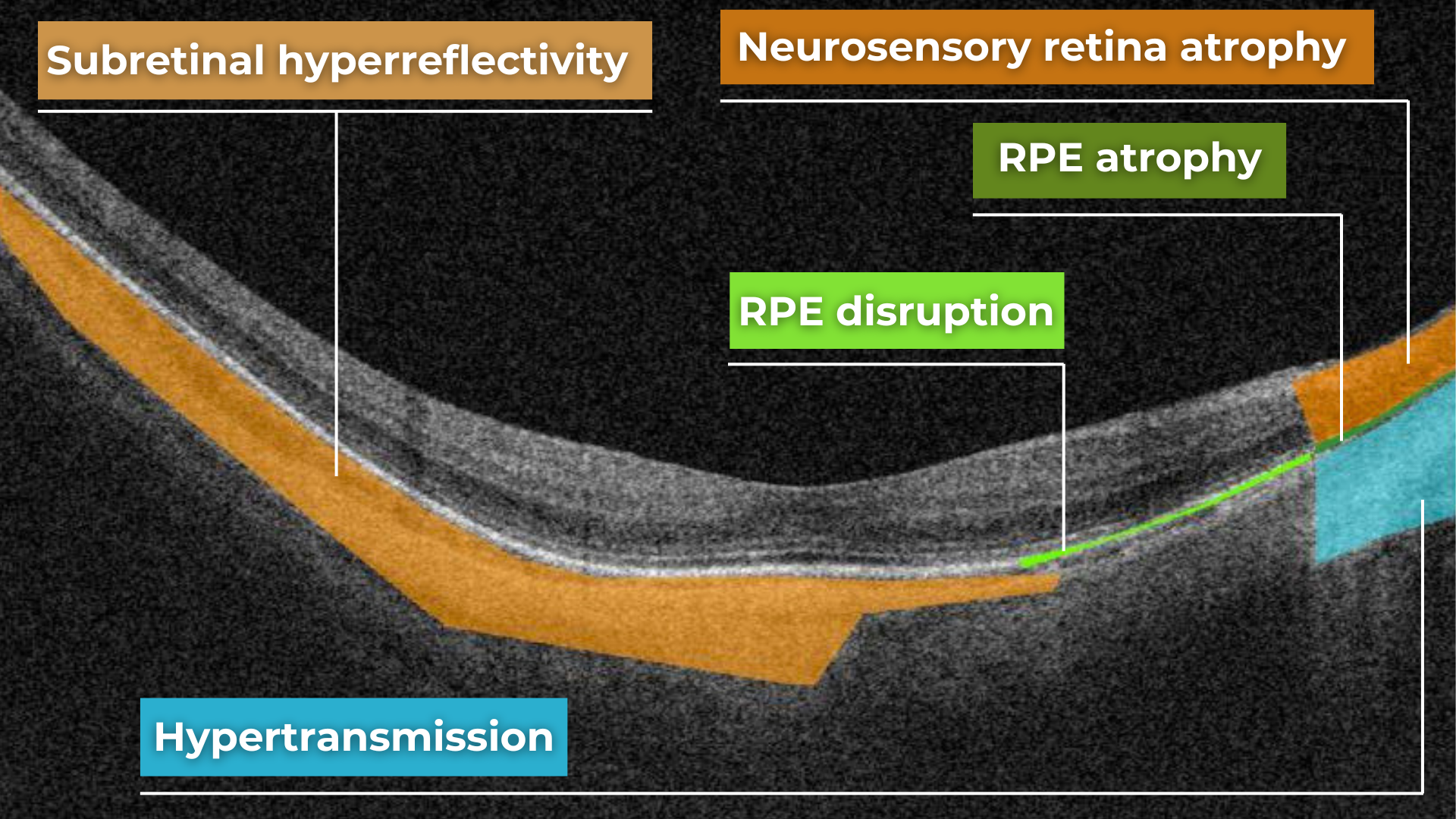
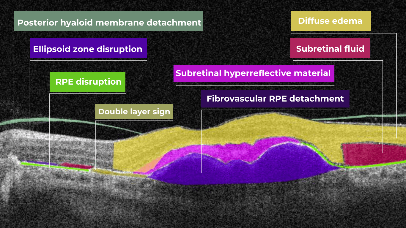
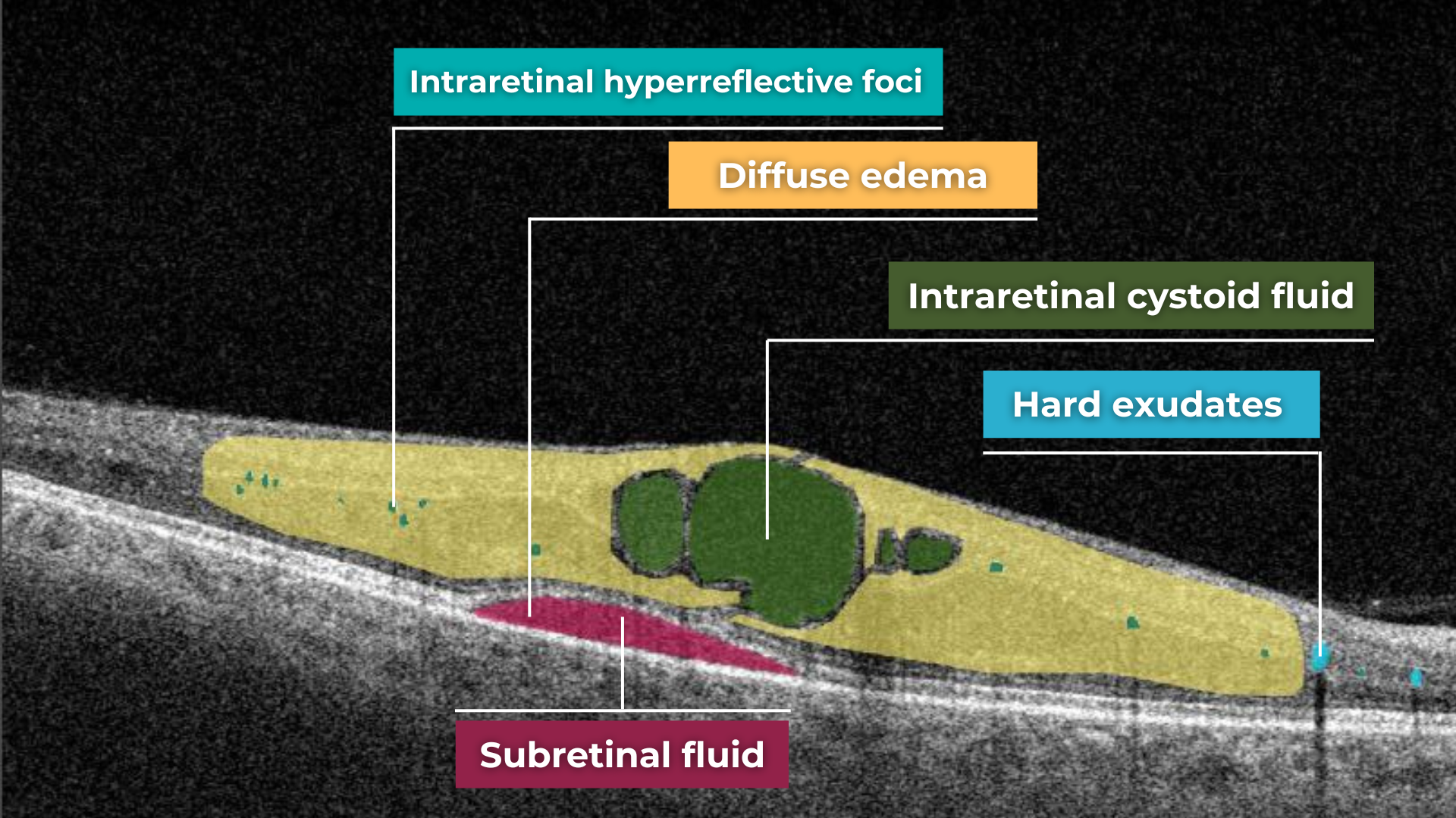
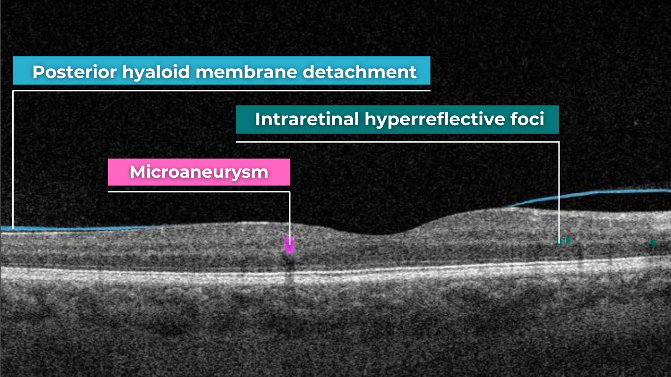
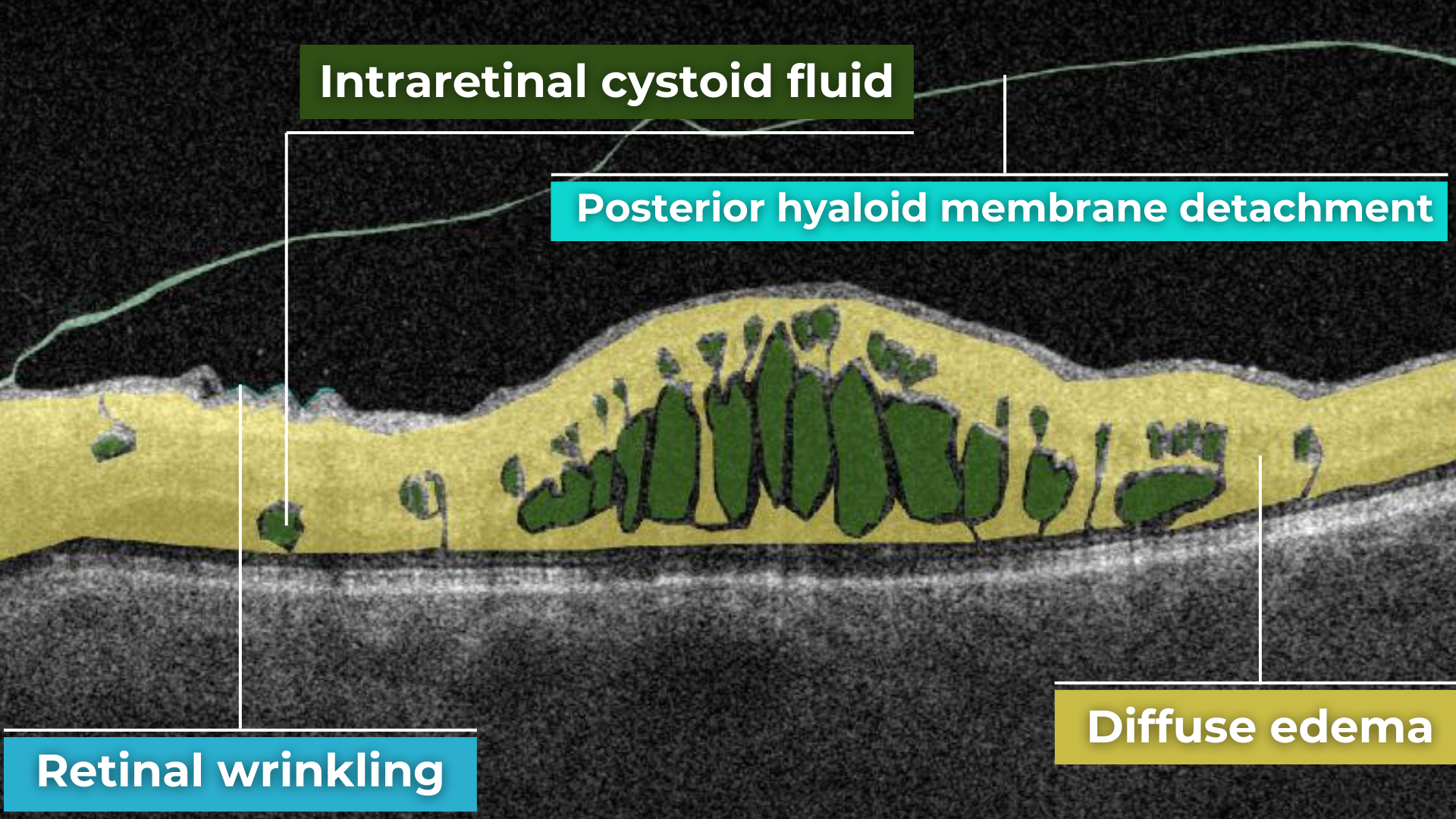

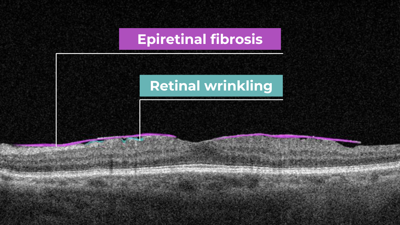

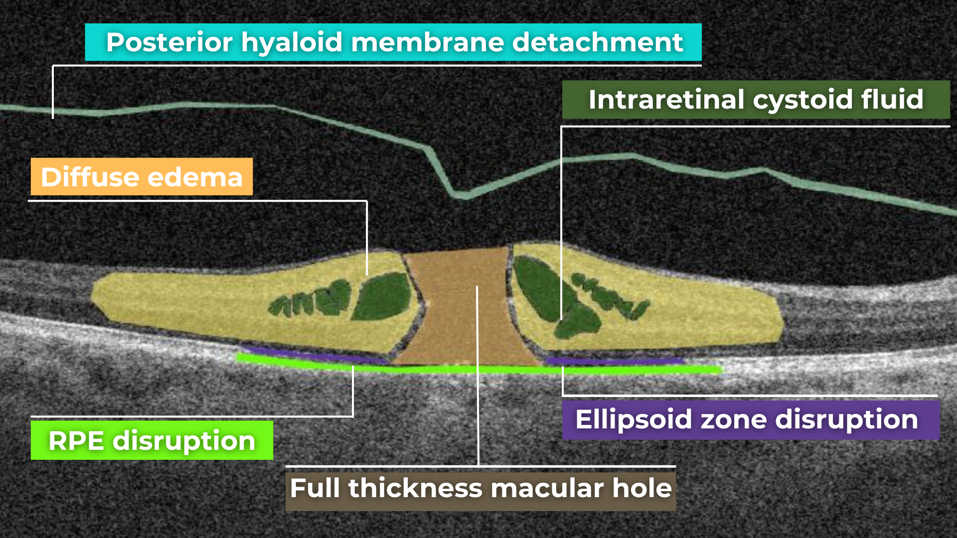
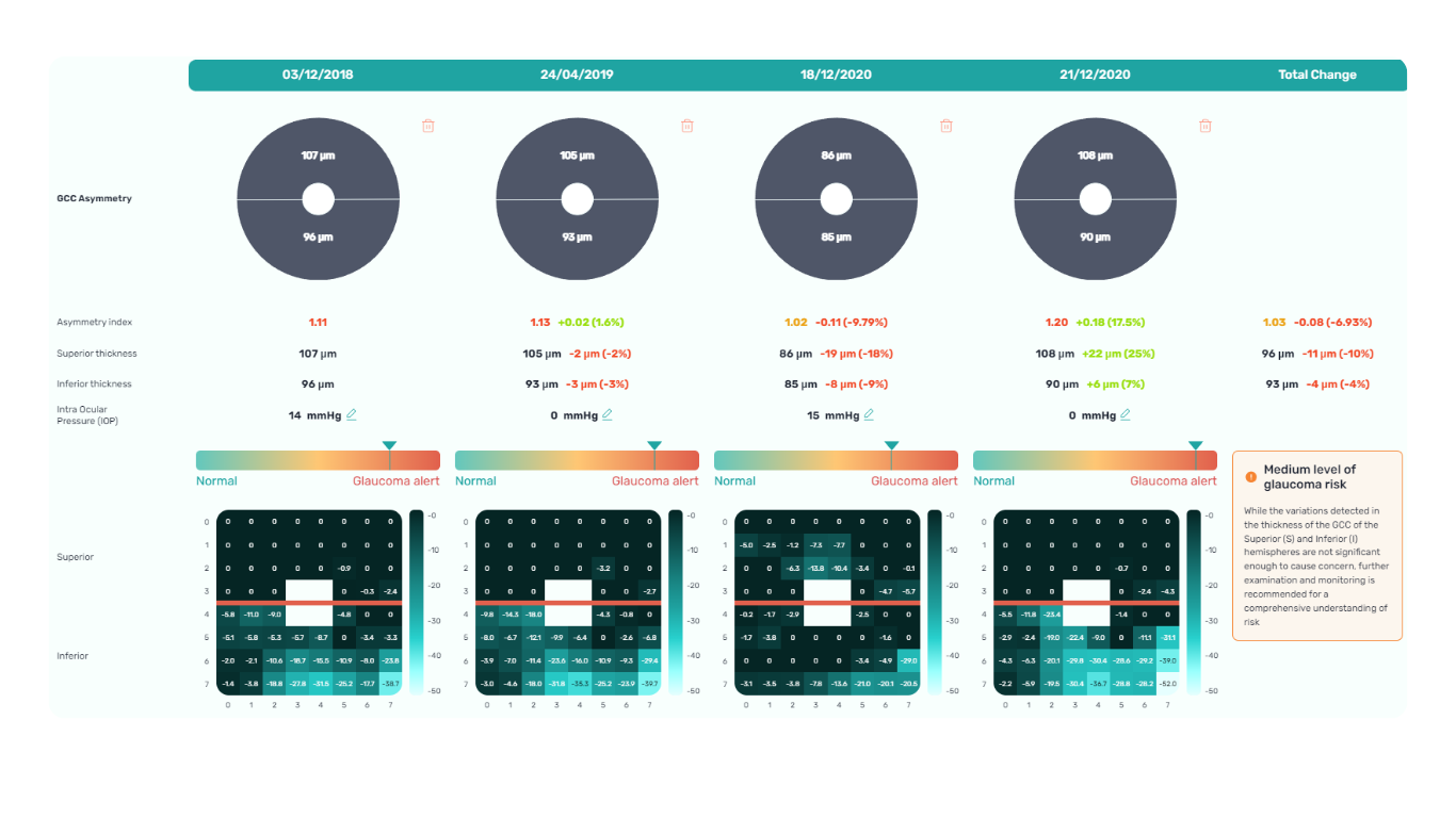
Recently Posted
-

Eye Hospital Management Software: Top 8 Solutions for your Clinic
Maria Znamenska
04.01.202310 min readThe term “Eye hospital management software” can have numerous meanings. Some soft can be a part of larger EMR (electronic medical records) systems, some can help with scheduling and billing, and some can help with patients’ information management. There is also an eye clinic management system that can even advise on diagnosis based on the patient’s history and medical images. Because of dozens of different soft on the market, it can be quite complicated to choose a proper set of tools for your practice.
If you are an ophthalmologist or manage an ophthalmic diagnostic center/hospital, you may have trouble choosing the right software. That is why we’ve decided to prepare a list of solutions for patients’ health recording and diagnosis. We will highlight the benefits of the ophthalmic practice management system and help you choose the right solution.
AI Decision Support for OCT
Why eye hospital management software is worth using
Eye hospital management software has become extremely important for eye clinics or medical centers looking to streamline their workflows, automate processes, and provide higher-quality care with less effort. You can have piles of paper and numerous excels, but when someone is on vacation, it will be impossible to make sense of all data and use it quickly.
However, many clinics still work according to the old scheme and refuse to introduce new technologies into their clinical practice. There may be several reasons for this: mistrust of modern tools, reluctance to spend the money buying licensed eye clinic management system, or reluctance to spend staff time learning how to work with the program. But, in fact, today, there are systems designed specifically for ophthalmologists to function flawlessly in eye care settings. Here are some benefits that an eye clinic management system can provide to your medical practice. Let’s take a closer look at some of them:

- High level of data protection. Another important benefit of the ophthalmic practice management system is a high level of data protection. High-quality soft gives access to data only to authorized persons. The software also has security systems that guarantee no risk of data loss and full protection of medical history or information about the patient’s condition.
- Increasing diagnostic accuracy. Using an eye clinic management system, ophthalmologists improve the quality of diagnosis and treatment, as they get access to the whole patient’s history from the past to the present. An ophthalmologist can learn about the previous treatment their patient received and about chronic illnesses. By learning this, doctors can create a better treatment plan.
- Increased revenue. Depending on the number of employees in your clinic, you may need dozens to hundreds of personnel to smoothly handle manual processes. And more human resources mean more expenses. However, by using best practice management software for ophthalmology, you can significantly reduce spending and let your employees and doctors focus on the more creative tasks that require empathy and communication.
These are the most common benefits of an eye clinic management system. However, each system has its unique features, so let’s look at the top 8 eye clinic management systems.
Altris AI System
Altris AI is a unique eye clinic management system that allows eye care specialists to analyze OCT scans with the help of artificial intelligence (AI) tools.
How does it work? Putting it simply, retina specialists have colored thousands of OCT scans and named more than 100 retinal pathologies and pathological signs to train an algorithm, so it can assist specialists in detecting the disease. After loading an OCT scan in the eye hospital management software, the AI model evaluates the b-scans (up to 512) and differentiates between normal scans and scans with moderate and severe pathology. It gives eye care professionals the ability to focus only on serious (red) scans, saving patients valuable time.
In addition, Altris AI allows its users to see a broader perspective of a patient’s eye health. All the reports are dynamically editable: the ophthalmologist can add/revise/delete items in the OCT report. Eye care specialists also can add segmentation/classification results to the OCT report in 1 click. And what’s even more important, Altris AI OCT report is understandable for both ophthalmologists and patients.
AI Decision Support for OCT
Eye clinic management system features of Altris AI
- The system allows working with all popular OCT equipment and all data storage formats, including DICOM of various lengths, png, and jpg.
- Altris AI ophthalmic practice management system can be easily integrated with EHR systems or run standalone as a web application.
- The system also takes care of user security, as all important patient data is tokenized and protected from disclosure at all stages.
- The artificial intelligence program can independently identify more than 100 retinal pathologies and pathological signs.
- The Smart Reports feature allows ophthalmologists to select the elements (single scan, layers, both eyes, etc.) that they want to see in their OCT report.
- This All Scans feature allows the user to view all scans of a single OCT examination, sort them by severity level, and zoom.
Watch a short overview of how Altris AI assists eye care specialists with OCT diagnosis and decision-making.
DrChrono Software

DrChrono EHR is an iPad and iPhone-compatible platform that offers fully customizable form templates or ready-made forms to help users track patient information.
DrChrono EHR is an iPad and iPhone-compatible platform that offers fully customizable form templates or ready-made forms to help users track patient information.
Eye clinic management system features of DrChrono Software
- The system allows medical practices to manage patient admissions, patient care, clinical charts, and billing.
- Healthcare professionals can add patient notes to the medical record. The Vital Flowsheets module provides the ability to create basic health data and monitor the health indicators of each patient.
- The DrChrono eye hospital management software also offers a variety of application integrations.
- Doctors can use the Free Draw module to annotate charts, OCT scans, or other files.
RXNT Software
RXNT is a comprehensive billing, practice management, and EHR solution. This system improves patient care and simplifies clinical management. Access patient health history and prescriptions at the point of care, schedule patients and providers, and request and review lab or imaging orders with multi-site single sign-on (SSO).
Eye clinic management system features of RXNT Software
- Any RXNT ophthalmic practice management system products (EHR, ERX, PM, Billing, Scheduling) can be combined into a fully integrated “Full Suite” system.
- Ophthalmologists, managers, or staff can add and organize documents in patient charts for clinical care plans and follow-up.
- The system has developed customizable “smart” forms and short keys that improve work processes.
- RXNT can share real-time data with other doctors to better coordinate care and support.
In addition, an ophthalmic clinic can integrate RXNT eye hospital management software with the Altris AI system to create and dynamically edit OCT reports.
Medfiles Software
Medfiles Software is a multi-task cloud-based solution that ensures compliance for ophthalmology clinic employees. The key features of this eye hospital management software are drug screening management, medical record tracking, case management, training tools, reporting, and safety documentation.
Eye clinic management system features of Medfiles Software
- Medfiles tracks patient treatment plans, open cases, treatment plans, medical expenses, and cash reserves and creates conclusions based on all the information.
- The system can be easily integrated with different software so a doctor or staff can see scans of specific OCT examinations.
Medfiles eye clinic management system allows to compare annual summary reports with benchmarks.
IntelleChartPRO Software
Another cloud-based ophthalmic electronic medical record (EMR) solution is IntelleChartPRO. This system is very popular among ophthalmology clinics and centers. IntelleChartPRO helps professionals record and manage a patient’s treatment and medical history more effectively.
Eye clinic management system features of IntelleChartPRO Software
- Physicians or ophthalmology clinic management can customize the EHR themselves to fit their unique workflows.
- IntelleChartPRO eye hospital management software developed adaptive template technology that allows offices to generate templates for each patient.
- In combination with other eye clinic management system tools, the software becomes more relevant and allows more accurate diagnoses of patients and the creation of detailed reports.
MaximEyes Software
MaximEyes is a comprehensive, unified electronic health record (EHR) and practices management solution designed exclusively for ophthalmology practices. It has a modern and intuitive user interface. The system will work on any computer OS. If users do not want to use cloud technologies or the clinic has a weak Internet connection, MaximEyes can be deployed through a local server
Eye clinic management system features of MaximEyes Software
- For each patient, the system allows ophthalmologists to set up an individual template according to different types of visits.
- The eye hospital management software EHR includes a flexible rules engine that will suggest or automatically generate post-diagnosis codes, procedure codes, and output documents.
- The First Insight module also offers an ophthalmic imaging management solution that works with any EHR.
75health Software
One more fully-fledged eye clinic management system is 75health, which is also a cloud-based solution that provides its users with electronic health record tools. 75health system will be most suitable for managing health records and patient information for ophthalmologists working in small and mid-sized clinics.
Eye clinic management system features of 75health Software
- 75health eye hospital management software allows ophthalmic clinic staff to download and save patients’ medical images, such as consent forms, handouts, or scans.
- Doctors can also create a treatment plan for their patients right in the system and scan records for allergies, medications, lab results, and symptom lists.
- 75health solution provides smooth integration of ophthalmic management systems, which helps ophthalmologists in decision-making.
myCare Integrity Software
Another cloud-based eye hospital management software that is worth your attention is myCare Integrity. It was created specifically for eye care specialists and contains a strong set of tools and modules that can cover the needs of any member of the ophthalmic clinic staff: from doctors to managers.
Eye clinic management system features of myCare Integrity Software
- The myCare Integrity system has an IntegriVIEW functionality that allows practitioners to link medical images directly to every screen of EMR.
- There is also an IntegriDRAW module inside the eye clinic software, where templates are included in the application. It allows users to rely on the previously created stamps.
- The IntegriLINK module allows ophthalmologists to link the diagnostic equipment to the system.
- What is more, myCare Integrity eye hospital management software allows you to customize and personalize the dashboard.
Summing up
Eye hospital management software is extremely important for any clinic, whether there are 10 or 500 employees. It can help you improve your workflow by keeping a lot of data in one place. Imagine how easily you can get rid of unnecessary paperwork, forget about administrative costs, and speed up processing. In addition, with an ophthalmic practice management system, you can get 24/7 access to patients’ data.
AI Decision Support for OCT
However, the key benefit of practice management software for opticians is the improvement of diagnosis and treatment. There are already ophthalmic image management systems, like Altris AI, that can not only help to manage patients’ data but also provide a second opinion regarding medical image analysis. Using this knowledge, doctors can have better access to patients’ health problems and reports, ultimately enabling them to provide the best care to their clients.
-
The Application of Machine Learning in Ophthalmology: The View from the Tech Side
Philip Marchenko
30.11.202215 min readAccording to the World Health Organization (WHO), artificial intelligence (AI) and machine learning (ML) will improve health outcomes by 2025. There are numerous digital technologies that shape the health of the future, yet AI and machine learning in ophthalmology and medical image analysis look like one of the most promising innovations.
The healthcare industry produces millions of medical images: MRI, CT, OCT, images from the lab, etc. The right diagnosis depends on the accuracy of the analysis by the specialists. Today AI can back up any medical specialist in medical image analysis: providing confidence and much-needed second opinion.
Try Altris AI for free
Check how artificial intelligence assists in OCT interpretation
Altris AI team decided to improve medical image analysis for just one type of medical image: Optical Coherence Tomography scans of the retina. To do it, the Altris AI team collected thousands of OCT scans and graphically labeled them, defining more than 100 pathologies and pathological signs. Watch the video to discover more features of Altris AI platform.
Then all this data was fed into the AI model. Further, I will tell how exactly we train the AI model of Altris AI so that it can detect more than 100 pathologies with 91% accuracy, but first, let’s discuss why it is important for the healthcare industry.
Why are automation and machine learning in ophthalmology important?
Due to the delicate anatomy of the eye, its treatment carries a high risk of complications. Sometimes these complications can be the result of a medical error by an eye care specialist. But how often?
According to the Altris team research, 20.2% of eye care practitioners miss minor, early, and rare pathologies on OCT scans 1- 3 times a week, and 4.4% miss them 3-5 times a week. But the worst thing is that 30.5% of ophthalmologists and optometrists are not even sure if they are missing any pathology at all.

Some medical errors may be minor, but some may cause significant harm to patients. Such medical errors can lead to medical malpractice lawsuits. That is why most ophthalmic clinics consider implementing AI to double-check the diagnosis of the ophthalmologist.
Besides, different tools of machine learning in ophthalmology have a high level of accuracy and can provide eye care specialists with a second opinion.
How to reach a high level of accuracy?
It is almost always necessary to conduct many experiments to achieve a high level of model accuracy (in the case of Altris AI, it is 91%). It is often done with the help of a machine learning pipeline.

High level of ML pipeline accuracy
The machine learning pipeline is programmed by a team of engineers to perform certain steps automatically. It systematically trains and evaluates models, monitors experiments, and works with datasets.
-
- ML and Medical teams collect, annotate and preprocess data. It’s crucial to ensure the data quality is at its highest level because the model’s quality heavily depends on it. To do this, the teams developed a process and annotation guideline, which ensures that the number of errors in the annotation is minimized.
- ML team chooses the appropriate approach (model) depending on the collected data and the tasks. Each team member is well-versed in the most modern and high-quality approaches that solve emerging tasks.
- The selected model is trained on the annotated data.
- In the model evaluating and testing stage, we develop tests aimed at helping us understand whether the model is trained properly to perform the needed tasks.
- After the ML team is satisfied with the result, we deploy the model, which means the model is ready for production.
- While the model is running in production, we monitor its performance to ensure everything goes well.
This workflow allows engineers to continuously fine-tune existing models alongside constant performance evaluations. The most significant advantage of this process is that it can be automated with the help of available tools.
Try Altris AI for free
Check how artificial intelligence assists in OCT interpretation
What tasks does machine learning in ophthalmology have?
Within the Altris AI platform, we solve 2 main tasks: segmentation and classification of OCT scans.
Classification task
Classification is the task of determining which category a particular object belongs to. We assign each pathology to a certain class of pathologies (for example, glaucoma class).
Segmentation task
The image segmentation problem can be stated as the division of an image into regions that separate different objects from each other, and from the background.
Key metrics of Altris ML pipeline
When discussing classification and segmentation metrics in medical imaging machine learning, it is essential to mention the Confusion matrix (CM). CM is a visualization of our performance, which helps us understand whether the model is performing well in terms of predicted and real data. For a better explanation, let’s take a look at the picture.
Let’s consider 4 possible outcomes from model predictions. Say we need to create a classifier to diagnose or predict if a patient has a disease (positive / 1 or TRUE) or not (negative/ 0 or FALSE). In such a case, the model can predict “yes” or “no”, and we can have an actual “yes” or “no”. Based on this, we can get 4 categories of results:
- TP — true positive. The patient that actually has a disease has been diagnosed with this disease. A class was predicted to be true, and it is actually true.
- TN — true negative. The patient is actually healthy and has been diagnosed as healthy. A class was predicted to be false, and it is actually false.
- FP — false positive (type 1 error). The patient that is actually healthy has been diagnosed as having a disease. A class was predicted to be true, but it is actually false.
- FN — false negative (type 2 error). The patient that actually has a disease has been diagnosed as healthy. A class was predicted to be false, but it is actually true.
With the help of the confusion matrix, our ML engineers get specific metrics needed to train our medical imaging machine learning model properly. We discuss each metric in more detail in the following paragraphs.
Classification metrics
- Accuracy
To find out how many of our predictions were correct, we divide the number of correct predictions by the total.

While being intuitive, the accuracy metric heavily relies on data specifics. If the dataset is imbalanced (the classes in a dataset are presented unevenly), we won’t get trustful results.
For example, if we have a training dataset with 98% samples of class A (healthy patients) and only 2% samples of class B (sick patients). The medical imaging machine learning model can easily give you 98% training accuracy by predicting that every patient is healthy, even if they have a disease. Such results may have destructive consequences as people won’t get needed medical treatment.
- Precision
Precision shows what proportion out of all positive predictions was correct.
Precision metric helps us in cases when we need to avoid False Negatives but can’t ignore False Positives. A typical example of this is a spam detector model. As engineers, we would be satisfied if the model sent a couple of spam letters to the inbox. However, sending an important non-spam letter to the spam folder (False Positive) is much worse.
- Sensitivity/Recall
Recall shows how many of all really sick patients we predicted and diagnosed correctly. It is a proportion of correctly positive predictions out of all positives.

In our case, you want to find all sick people, so it would not be so critical if the model diagnoses some healthy people as unhealthy. They would probably be sent to take some extra tests, which is annoying but not critical. But it’s much worse if the model diagnoses sick people as healthy and leaves them without treatment.
The sensitivity of Altris AI is 92,51%
- Specificity
The specificity shows how many of all healthy patients we predicted correctly. It is the proportion of actual negatives that the medical imaging machine learning model has correctly identified as such out of all negatives.
Specificity should be the metric of choice if you must cover all true negatives and you can’t tolerate any false positives as a result. For example, we’re making a fraud detector model in which all people whose credit card has been flagged as fraudulent (positive) will immediately go to jail. You don’t want to put innocent people behind bars, meaning false positives here are unacceptable.
The specificity of Altris AI is 99,80%
Segmentation metrics
Segmentation also can be thought of as a classification task. For each pixel, we make predictions about whether it is a certain object or not. Therefore, we can talk about Accuracy, Precision, Recall, and Specificity in terms of segmentation.
Let’s say we have a Ground Truth (what is really an object) and a Segmented (what the model predicted). The intersection in the picture below is the correct operation of the medical imaging machine learning model. All that is the difference (FN and FP) is the incorrect operation of the model. True negative (TN) is everything the model did not mark in this case.
Quite often, even after looking at such metrics, the problem of non-symmetricity remains in the segmentation tasks. For example, if we consider a tiny object, the Accuracy metric doesn’t work. Therefore, segmentation tasks also refer to additional metrics that allow taking into account the size of the object of the overall quality assessment. Let’s look at additional metrics in more detail.
- Intersection over Union (IoU)/Jaccard
Intersection over Union is an evaluation metric used to measure segmentation accuracy on a particular image. This metric is considered quite simple — the intersection zone is divided by the union of Ground Truth and Segmented.
Sometimes we get such results, like if the object was determined to be very large, but in fact, we see that it is small. Then the metric will be low, and vice versa. If the masks are approximately equal to each other, everything works correctly, and the metric will be high.
- Dice score/F1
The dice coefficient is 2 times the area of overlap divided by the total number of pixels in both images.
This metric is a slight modification of the previous one. The difference is that, in this case, we take the intersection area twice.
Calculating scores over dataset
We calculate the metrics described above for each scan. In order to count them over the entire dataset, we take each picture in this dataset, segment it, calculate the metric, and then take the average value of the metrics on each image.
What is model validation in ML?
In addition to evaluating the metrics, we also need to design the model validation procedure suitable for a specific task.
When we have determined the metric that suits the task of machine learning for medical image analysis, we also need to understand what data to use for calculation. It will be wrong to calculate the metric on the training data because the model has already seen it. This means that we will not check the ability of the model to generalize in any way. Thus, we need a specific test dataset so that we can carry out quality control according to the selected metrics.
The main tasks of the model validation are:
- To provide an unbiased estimation of the accuracy that the model can achieve
- To check whether the model is not overfitted
Picking the correct model validation process is critical to guarantee the exactness of the validation method. In addition, there is no single suitable validation method for machine learning in ophthalmology — each task requires different validation. Engineers separately examine each task to see if data has leaked from the train dataset to the test dataset because this may lead to an overly optimistic estimate of the metrics.
For example, we can take OCT images in different resolutions. We may need a higher image resolution for some diseases. If the medical imaging machine learning model overfits at the resolution of this OCT, it will be called a leak because the model should behave the same at any resolution.
Overfitting and underfitting
The model also has such an important property as a generalization. If the model did not see some data during training, it should not be difficult for the model to determine which class a certain image belongs to.
At this stage, engineers may have two problems that they need to solve. The first problem is overfitting. When the model remembers the training data too well, we lose the ability to make correct predictions. The picture below illustrates this problem. The chart in the middle is a good fit when the model is general enough and has a positive trend, and the trend is well-learned. But the chart on the right shows a too-specific model that will not be able to guess the trend.
Another problem to solve is underfitting. This problem arises when we have chosen a model that is not complex enough to describe the trend in the data (left chart).
Bias variance trade-off
Another important concept we use in machine learning model validation is the bias variance trade-off. We want our models to always make accurate predictions and have no ground truth scattered. As shown at first/second circle.
However, there are situations when we have a model that predicts something close to the target, but from dataset to dataset, it has a strong scatter. This is showcased in the second circle.
In circle three, you can observe a situation where the model has heap predictions on different datasets, but they are inaccurate. This situation usually indicates that we need to almost entirely rebuild the model.
Overfitting and bias variance trade-off are very important in working with the model, as they allow us to track errors and select a model that will balance between spread and hitting the target.
Unbiased estimation
In addition, within each model, we evaluate a set of parameters. We made a certain estimate (graph on the left), but in real life, the distribution of parameters differs (graph on the right). Thus, seeing that our estimate turned out to be shifted, we find another problem that needs to be solved. Machine learning in ophthalmology needs to make the estimate as unbiased as possible.
How do we validate the Altris AI model?
There are three main steps in choosing a validation strategy for machine learning in ophthalmology:
- we got familiar with ophthalmology, understood the nature of data, and where the leakages are possible;
- We estimated the dataset size and target distribution;
- understood the model’s training complexity (amount of operations/ number of parameters/ time) to pick the validation algorithms.
After that, we have a reliable strategy for the machine learning model validation. Here are some fundamental concepts we use in the validation of models’ performance.
Train/test split
Train/test split is the most simple and basic strategy that we use to evaluate the model quality. This strategy splits the data into train and test and is used on small datasets. For example, we have a dataset of 1000 pictures, 700 of which we leave for training and 300 we take for the test.
This method is good enough for prototyping. However, we don’t have enough datasets with it to do a simple double-check. This phenomenon is called high sampling bias: this happens when we encounter some kind of systematic error that did not fit into the distribution in the train or test.
By dividing data into train and test, we are trying to simulate how the model works in the real world. But if we randomly split the data into train and test, our test sample will be far from the real one. This can be corrected by constructing several test samples from the number of data we have and examining the model performance.
Train/test/holdout set
We leave the holdout as the final validation and use the train and test to work with the medical imaging machine learning model. After optimizing our model on the train/test split, we can check if we didn’t overfit it by validating our holdout set.
Using a holdout as a final sample helps us look at multiple test data distributions and see how much the models will differ.
K-fold cross validation
There is also a more general approach that Altris AI team use for validation — k-fold cross validation. This method divides all of our data equally into train and test.
We take the first part of the data and declare it as a test, then the second, and so on. Thus, we can train the model on each such division and see how it performs. We look at the variance and standard deviation of the resulting folds as it will give information about the stability of the model across different data inputs.
Do we need ML models to perform on par with doctors?
Here I will try to answer a question that worries many ophthalmologists and optometrists: can machine learning for medical image analysis surpasses an eye care specialist in assessing quality?
In the diagram below, I have drawn an asymptote called the Best possible accuracy that can be achieved in solving a particular problem. We also have a Human level performance (HLP), which represents how a person can solve this problem.
HLP is the benchmark that the ML model strives for. Unlike the Best possible accuracy, for which there is no formula, HLP can be easily calculated. Therefore, we assume that if a model crosses the human quality level, we have already achieved the best possible quality for that model. Accordingly, we can try to approximate the Best possible accuracy with the HLP metric. And depending on this, we understand whether our model performs better or worse.
For those tasks that people do better and the ML model does worse, we do the following:
- collect more data
- run manual error analysis
- do better bias/variance analysis
But when the model crosses the HLP quality level, it is not entirely clear what to do next with the model and how to evaluate it further. So, in reality, we don’t need the model to outperform a human in interpreting images. We simply won’t know how to judge the quality of this model and whether it can be 100% objective and unbiased.
Avoidable bias
Let’s say we need to build a classifier for diabetic retinopathy based on OCT scans, and we have a control dataset prepared by people. In the first case, doctors are wrong 5% of the time. At the same time, the model on the train set is wrong in 10% of cases and on the test set — in 13%.
The difference between the model’s and the human’s performance is usually taken as the minimum difference between the train/test set and the human. In our case, it is 5% gap (10% – 5%) of avoidable bias. It is called avoidable bias because it can be fixed theoretically. In such a case, we need to take a more complex model and more data to better train the model.
In the second case, doctors determine the disease with a 9% error. If the model defines a disease with the same rates as the previous example, then the difference between the train/test set and the human will be 1% (10% – 9%), which is much better than avoidable bias.
Looking at these two cases, we must choose a strategy that will lower the variance for the machine learning model so that it works stably on different test sets. Thus, taking into account the avoidable bias and the variance between the samples, we can build a strategy for training the model so that it could potentially outperform the HLP someday. However, do we need it now?
Understanding HLP
To better understand the HLP metric, let’s consider the task of determining dry AMD on OCT scans. We have a fixed dataset and 4 train sets, each one determining dry AMD with a specific accuracy:
- ML engineers – 20%
- ophthalmologists – 5%
- 2 ophthalmologists – 3%
- 2 ophthalmologists and 1 professor of ophthalmology – 2%
We take a result of 2% as the best HLP possible. To develop our model, we can choose the performances we strive to get. The 20% error result is irrelevant, so we discard this option. But the level of 1 doctor is enough for model version number 1 model. Thus, we are building a development strategy for model 1.
Summing up
Machine learning will revolutionize the eye care industry. It provides confidence and second opinion to eye care specialists in medical image analysis.
If you are looking for ways to use machine learning in your eye care practice, feel free to contact us. At Altris AI, we improve the diagnostic process for eye care practitioners by automating the detection of 54 pathological signs and 49 pathologies on OCT images.
-
-
Altris AI Builds Partnership with Academic Institutions
Maria Znamenska
21.11.20222 min readAI in ophthalmology for academic purposes
Aston University and Altris AI join forces to Revolutionise Optometry Education
We are proud to announce our new strategic partnership with Aston University, a renowned healthcare education and research institution. This partnership marks a significant step forward in enhancing the preparation and training of optometry students with the help of AI technology.
As OCT examination proves to be one of the most accurate and yet the most complex diagnostic devices in the eye care industry, it is crucial for educational institutions to stay ahead and equip their students with the most innovative tools, such as artificial intelligence. The collaboration between ourselves and Aston University will enhance how optometry students learn and improve OCT interpretation skills.
Aston University, known for its commitment to excellence in healthcare education, has chosen to partner with Altris AI to integrate AI-driven solutions into future optometrists training in the lecture theatre, university clinics, and research departments. The Altris AI platform will also be used in the study of Ph.D.-level research projects.
Commenting on this exciting partnership, James Wolffsohn, Head School of Optometry said “We strive to equip our students with cutting edge knowledge and tools to deliver world class eyecare to their patients. This partnership with Altris AI will help strengthen our students diagnostic ability and keep on the crest of the innovation wave offered by AI”
Maria Znamenska, MD, Ph.D., Associate Professor of Ophthalmology and a Chief Medical Officer at Altris AI, expressed her enthusiasm, stating, “The new generation of optometry students ask for modern ways of learning. Today they want more than books and atlases, they want to learn interactively and utilise the power of technology in clinical practice. And we are happy that AI has become a true copilot for the young generation of optometry students at Aston University.”
This partnership is a testament to the commitment of both organisations to innovation in healthcare education. Together, Aston University and Altris AI aim to shape the future of optometry education and empower students to provide excellent level eye care services to patients. After all, the ultimate goal of digitalisation in healthcare is always healthier patients.
Contact us
Ask us any question -

Will Artificial Intelligence Replace Ophthalmologists & Optometrists?
Maria Znamenska
17.11.20228 min readWill optometrists be replaced by AI?
Back in 2019, at the World Science Congress, Peter van Wijngaarden, Deputy Director of the Center for Eye Research, claimed that the eye sector is one of the leading areas of medicine in terms of artificial intelligence (AI) implementation. According to RANZCO, AI systems are already achieving incredible results and, in some cases, can even rival eye care specialists.
Since AI has become a buzzword, there are hundreds of articles, speeches, and videos on this topic. We are the company that created AI for the eye care and we know the answer to this question: ” Will AI take over optometry and ophthalmology?” ( Spoiler: NO).
Try a co-pilot AI for OCT analysis ( but it won't replace you)
It is simple a misconception. There are a lot of similar examples of AI misconceptions when famous professors and specialists in the field of ophthalmology made predictions that artificial intelligence is rapidly gaining strength in the eyecare industry. This gives rise to many myths and fears around the introduction of AI in clinical practice. Will optometrists be replaced by AI? What about ophthalmologists? What is going on?
The increased attention to the issue of optometrists and ophthalmologists replaced by AI was also provoked by a World Economic Forum (WEF) report. According to this report, people can lose 85 million jobs by 2025 due to the shifting division of labor between people and machines.
In this post, we will discuss the top 5 AI misconceptions that are most often faced by the owners of ophthalmological clinics and optometry centers in order to dispel them once and for all.
What exactly is AI? Do AI algorithms work exactly like a human brain?

The concept of optometrists and ophthalmologists replaced by robots is gaining popularity. Nowadays, eye care specialists often discuss the potential of AI training in human cognitive skills. It is no longer just about the ability of AI to detect Diabetic retinopathy or interpret OCT scans with greater accuracy. The question is, will AI ever be able to replicate human consciousness? And can AI replicate how the human brain works?
What do we know about such models in different areas? AI systems are already demonstrating the work of some human cognitive functions. For example, AI models successfully compete with humans in computer games by gradually learning successful strategies. There is also an AI model which creates enjoyable melodic music.
However, replacing optometrists and ophthalmologists with AI still seems VERY unrealistic. Even with the above examples mimicking some aspects of human behavior, an AI algorithm still needs to learn what empathy is. Artificial intelligence does not understand and cannot make sense of its surroundings, nor can it learn from its surroundings as humans do. The most famous example that confirms this inability of AI is Siri or Alexa. Voice assistants can set up appointments but give strange answers when the conversation goes differently than their scenario.
While the human brain inspires modern AI techniques such as neural networks (NNs), the structure of NNs architectures is not biologically realistic.
First of all, there is a set of qualities that ophthalmologists and optometrists use every day. It is empathy for the patient, as well as creativity, teamwork, and adaptability. These qualities help doctors provide effective care to their patients. It is unlikely that the machines will ever be able to work with children, older adults, or patients with specific disabilities on par with humans. In addition, any patient would like to hear the diagnosis or discuss a treatment plan with a doctor, not a machine.
Therefore AI algorithm can’t work like a human brain, and the scenario where artificial intelligence replace ophthalmologists and optometrists will never happen. Nowadays, there are no developments that would make us think that AI image interpretation will ever be able at least to repeat important qualities of eye care specialists.
Is today’s state of AI dangerous for humans?
Today, AI algorithms can interpret retinal images and distinguish pathological from non-pathological scans. However, not all attempts at AI implementation have succeeded as well. One of the most popular non-medical examples is Facebook. Some time ago, Facebook tried to identify relevant news for certain groups of users. But the automated process could not detect the difference between real and fake news. Russian hackers managed to trick the system and bypass automatic filters. They posted fake news, forcing the Facebook team to come back to human editors.
This is just one example of how security lags behind performance when humans rely on AI too much. Artificial intelligence is a great tool, but in most cases, its abilities only give reliable and the most accurate results in collaboration with eye care professionals. Although machines are designed by humans, they often can’t predict human behavior and don’t know how to cope with situations or clinical cases that go beyond the scope of the algorithm.
Therefore AI is not dangerous for humans when ophthalmologists and optometrists periodically control the work of algorithms and review how the machine works. This is the number-two reason why artificial intelligence replace ophthalmologists and optometrists is unrealistic.
Will AI ever be 100% objective?
To honestly answer the questions of will artificial intelligence replace ophthalmologists and optometrists and whether it is 100% objective, you need to understand that an AI system will only be as good as its inputs. By loading unbiased training datasets, engineers can create an AI system that makes unbiased decisions. However, in the real world, AI is unlikely ever to be 100% objective.
For example, many well-known companies, such as Amazon or Facebook, still struggle with the gender gap in hiring. Some time ago, Amazon used historical data from the past ten years to train its AI recruiting model. The algorithm was supposed to process data and candidates and free recruiters from the routine viewing of hundreds of CVs. However, soon Amazon team discovered that the data was biased against women. AI algorithm was trained by outdated information when the technology industry used to be dominated by men. Thus, the new recruitment system selected only male candidates. This forced Amazon to abandon the algorithm and re-open many recruiter positions.
In the field of ophthalmology, AI models can already accurately predict diabetes risk factors or potential vision loss from OCT images. So when will artificial intelligence replace ophthalmologists? In Altris, we are sure the algorithm will never achieve adequate objectivity, as it will always be limited by input data, whether demographics, gender, or age.
Now we know that AI can’t be 100% objective. Indeed, ophthalmologists and optometrists can’t match the ability of algorithms to detect pixel-level patterns among the millions of pixels in the OCT scan. However, only the cooperation of eye care specialists and a quality AI model working together will allow for more accurate detection of diseases. The combined efforts of AI management systems and eye care specialists can help achieve the desired 100%.
Will optometrists be replaced by AI?What about ophthalmologists?
Various articles have speculated on whether artificial intelligence replace ophthalmologists and optometrists, raising concerns about unemployment. However, this never corresponded to the actual state of affairs. Carl Benedikt Frey, an Oxford Martin Citi Fellow at Oxford University, reported that while 47% of jobs are at risk of automation, the risk for doctors is estimated at only 0.4%.
In addition, in his book “Humans Are Underrated”, Geoff Colvin states that the most valuable skill for ophthalmologists is the ability to sense the thoughts and feelings of patients who are losing sight.
Many patients complain about the lack of contact with the doctor. They admit that the treatment would be more comfortable if doctors devoted more time to live communication. This mainly applies to children and the elderly, who need a lot of attention from eye care specialists. Empathy and similar human qualities are not only an understanding of the patient’s feelings but also an adequate response to them. Thus, a future in which optometrists and ophthalmologists are replaced by AI seems senseless.
Professor Tien Yin Wong, medical director of the Singapore National Eye Centre, claimed that AI holds great promise for retinal screening. And while AI for OCT interpretation will radically change clinical practice, the technology’s more significant impact will be to complement and enhance human capabilities rather than replace them. The field of ophthalmology demonstrates that the combined efforts of scientists and machines are more effective than either could achieve individually.
Artificial intelligence for OCT interpretation is just a recommendation system for an eye care specialist. Often one pathological sign, for example, Cystoid macular edema (CME), or Intraretinal fluid, can indicate many diseases, like Wet AMD, DR, DME, CRVO, and others. That is why AI is only an assistant to a doctor, especially when it comes to rare pathologies.
All in all, AI for OCT interpretation is just a tiny part of clinical practice and can never work without humans. In order to detect the pathological signs and diagnose a disease correctly, an eye care specialist must perform different examination methods. Among these exams are visual acuity, intraocular pressure, ophthalmoscopy, and a basic patient examination, which includes anamnesis. Moreover, ophthalmologists and optometrists may also need to perform other visualization methods, like Fundus photography, FFA, or OCTA.
Will AI replace optometrist?
This is probably one of the key AI misconceptions. Automation has led to a significant change in many industries, and ophthalmology is no exception. So when will AI take over optometry and ophthalmology? The answer is quite simple — AI will never replace eye care specialists. It will eventually take over routine tasks, allowing the careers of ophthalmologists and optometrists to advance in new and exciting directions.
Try a co-pilot AI for OCT analysis ( but it won't replace you)
Automated interpretation of OCT scans will significantly increase the circulation of patients in ophthalmic clinics or optometry centers, which is commercially attractive. Moreover, with increasing life expectancy, and expanding the range and effectiveness of treatment options offered, a collaborative effort between ophthalmologists and AI will improve patient outcomes. This will make ECPs more efficient, freeing up time for human interaction between doctor and patient, which has been a cornerstone of medicine for decades.
There are hundreds of eye care specialists who are already using AIf for OCT scan analysis, for example, to improve! the results. So will AI take over optometry or ophthalmology? The answer is rather simple: No
-
AI for Reading Centers: How it Boosts Workflow and Efficiency
Mark Braddon
05.10.20227 min readIn recent years reading centers have become an essential resource for facilitating imaging research in many fields, including clinical trials of ophthalmology drugs. And their importance will continue to grow.
Reading centers provide crucial information by evaluating images. That is why for conducting accurate clinical trials, they must hire ophthalmologists of high qualification. Moreover, to ensure consistent analysis, the materials that graders use for the research (be it fundus photographs, fluorescein angiograms, or OCT scans) must also undergo quality control. However, even such measures can’t completely exclude errors or biases.
Meanwhile, recent developments in the field of AI medical image analysis revolutionized the approach to clinical trials, which makes it possible to boost the workflow of reading centers. AI image analysis software works with thousands of images, efficiently providing the large amount of data needed to analyze the patient’s condition. In addition, evaluating images with AI is faster, cheaper, and more effective.
Book demo for a company
Try artificial intelligence for OCT analysis
In this article, we will discuss the top 5 benefits of AI medical image analysis software for reading centers and the way AI improves the image interpretation process.
Limitations of the manual evaluating procedure
Although several reading centers have already implemented AI for medical image analysis in their workflow, most organizations are far from evaluating automation and prefer classic image interpretation methods.
In most reading centers, ophthalmologists manually evaluate ocular images for drug safety studies, compile the images, and perform statistical analysis of the data. Research sizes for reading centers can range from 50 images to 3000 or more, and dozen of separate sets of images can be collected per research subject. Therefore reading centers have many obstacles to a quality evaluation process and accurate results.
-
Large amount of images is hard to proceed
The vast number of images that need to be processed in the short term usually leads to the main problem for reading centers — most hire outsourced ophthalmologists to speed up the image grading and evaluation process. Outsourced specialists have different levels of qualification and different evaluating methods, which may lead to decreased accuracy. In addition, outsourced eye care specialists are not always interested in performing the work at the highest level.
-
Human resources are expensive
Another limitation of the standard evaluating procedure is the high сost spent on ophthalmologists. Human resources are usually quite expensive and associated with the risk of staff turnover.
-
High probability of human bias
Besides, hours spent in front of a computer screen evaluating thousands of images create a stressful environment for ophthalmologists and cause many errors, affecting the accuracy of the clinical trials. Even the FDA recognizes grader fatigue and its impact on potential errors in image interpretation.
-
Inaccurate labeling
In addition, administrative problems also occur quite often. This happens due to deviations from study protocols and incorrect labeling of images, which can compromise the integrity of the analyses.
Fortunately, the pace of digitalization in reading centers is accelerating. Here is how AI medical image analysis can help reading centers cope with the growing workload.
The importance of implementing AI medical image analysis for reading centers
Usually, AI image analysis is made through a pattern recognition process that involves scanning images for specific pathological signs to interpret the patient’s condition. The AI image analysis software has precise and efficient evaluation protocols that allow the analysis and interpretation of images in terms of a variety of qualitative morphological parameters. For example, when analyzing images of a patient with diabetic retinopathy, the AI models recognize microaneurysms or hemorrhages.
AI algorithms allow reading centers to conduct trials of any size and duration, including various treatments for various eye diseases. Moreover, unlike the standard image interpretation process, which requires significant human resources, the introduction of AI for image analysis into the workflow of reading centers has many advantages.
- Quality control. Using AI algorithms ensures no errors in OCT scan analysis. AI image analysis software ensures that the desired parameters are classified based on certified imaging protocols.
- Less money spent. Implementing AI-assisted OCT analysis is less expensive than hiring outsourced ophthalmologists.
- Accurate quantification. AI in medical image analysis does not depend on patient characteristics or treatment group assignment knowledge, so the machine provides the most objective and accurate assessment possible.
- Increased efficiency. Improving the reading centers workflow with AI provides an objective and standardized classification of images. It means that any human bias is excluded, which increases the reputation of clinical research.
- No time wasted — no more hours spent at a computer screen. Evaluating images with AI medical image analysis provides faster and more sensitive identification of the patient’s condition, which can positively impact decision-making.
Book demo for a company
Try artificial intelligence for OCT analysis
How reading centers will benefit from AI image analysis software
In short, image evaluation with algorithms is fast, less expensive, and more reproducible. However, many companies that perform clinical trials in cooperation with reading centers are still afraid of implementing AI in medical image analysis and evaluating processes. Modern AI-based image management systems, such as Altris AI, unlike their predecessors, allow reading centers to overcome the challenges of the manual image interpretation process.
A lot of data available to train an algorithm
The more images with various pathological features the algorithm has for training, the more accurately it will detect the diagnosis. Modern AI image analysis software has the ability to obtain thousands of OCT images from different models of devices for comprehensive and correct training of algorithms. Although many medical centers keep their clinical practice confidential, many ophthalmic cases and images with various pathological signs in the public domain allow the training of AI algorithms.
For example, the Altris AI medical image analysis software was trained on 5 million unique OCT scans obtained in 11 practicing ophthalmology clinics through the years. Our retina experts took a responsible approach to annotating and labeling images for algorithm training. A thorough error detection and correction procedure gave our algorithm 91% accuracy.
Constant quality control
The responsibilities of the modern algorithms developers include not only the release of the model but also further diagnostics, which allows avoiding the problem of reproducibility. After all, constant quality control is necessary for algorithm development environments. Understanding the importance of quality control, the Altris AI team constantly tests the reproducibility of AI medical image analysis model diagnostics.
Collection of rare diseases
According to our research, 25% of ophthalmologists, on average, miss rare pathologies 3 times a week. However, modern AI image analysis software allows overcoming this challenge. For example, Altris AI excludes missing minor, early, rare pathologies. Our team created an algorithm that automates the detection of 54 pathological signs and 49 pathologies.
High percentage of algorithmic bias is avoided
Algorithmic bias is one of the biggest challenges in AI. Although algorithms themselves do not have biases, they inherit them from humans. However, today, AI for image analysis has learned how to overcome the lack of interoperability between medical record systems.
Although it is impossible to avoid algorithmic bias completely, as it can appear at any stage of the algorithm creation process, from study design and data collection to algorithm development and model selection, modern developers take a direction to fair AI. By using a technical and regulatory framework that provides the diverse data needed to train AI algorithms, the Altris AI team makes modern technologies inclusive and ensures algorithmic bias can be excluded.
The future of AI medical image analysis in reading centers
The ultimate goal of the ophthalmic AI system for reading centers is to improve the grading and evaluating process and obtain more accurate research results. However, instead of fully digitalizing image assessment, the ideal approach to analysis is integration — where the benefits of AI algorithms and human skills can be combined.
Technology will never fully replace humans, but it is already improving their work efficiency. For example, by taking over more routine and monotonous tasks, algorithms allow ophthalmologists to focus on specific eye areas and increase the evaluation speed. AI medical image analysis software can also be effective in determining compliance with the standardization of feature interpretation and determining image quality for requesting more images.
There are undoubtedly many challenges to integrating AI for image analysis into the workflow of reading centers. However, modern AI technologies can already overcome almost all of them. Altris AI image interpretation system is changing the future of clinical research by helping to classify images faster and increasing the efficiency, accuracy, and reproducibility of clinical trial data.
You can watch a short video of how Altris AI platform assists eye care specialists in detecting pathological signs on the OCT scans:
-
-
The Role of AI Image Interpretation for Ocular Pathologies Detection
Maria Znamenska
28.09.202220 min readThe burden of timely diagnostics lies on the shoulders of eye care specialists: ophthalmologists and optometrists worldwide. According to the International Agency for the Prevention of Blindness, over 1 billion people live with preventable blindness because they can’t access the proper diagnostics and treatment. Almost everyone needs access to eye care services during their lifetime. Unfortunately, there are only 331K optometrists worldwide, while 14M optometrists are required to provide effective and adequate eye care services.
With the high prevalence of the population that needs eye care services and the lack of specialists, the goal of timely and accurate diagnostics and treatment seems unachievable.
See how AI for OCT works
However, the empowerment of eye care specialists with Artificial Intelligence (AI) can be a real solution to this problem. As the larger part of the work of eye care specialists relies on retina image assessment and analysis, the support of this process can unburden ophthalmologists and optometrists all over the world. Modern AI image interpretation algorithms, such as Altris AI, can discover patterns among millions of pixels with high speed, accuracy, and zero human errors because of tiredness.
You can watch a short video of how Altris AI can assist you in detecting pathological signs on the OCT scans:
https://www.youtube.com/watch?v=Ehhwl6Q0O-A&ab_channel=Altris
In this article, we will talk about the capabilities of AI image interpretation for Optical Coherence Tomography (OCT) in detecting common pathologies, such as AMD or glaucoma, and less prevalent, such as Choroidal Melanoma. Despite the skepticism of the eye care community towards AI, multiple research works mentioned in this article prove the efficiency of AI. Moreover, there are market tools, capable of detecting 49 eye pathologies with 91% accumulative accuracy. Altris, the SaaS created by a team of retina experts based on 5 million OCT scans obtained in 11 clinics, is such a tool.
AI image interpretation for OCT
AI image interpretation for Asteroid Hyalosis
Asteroid hyalosis is a clinical condition in which calcium-lipid complexes are suspended throughout the vitreous collagen fibrils. Although it is a rare disease (1.2% prevalence according to the U.S. Beaver Dam Eye Study), it also may lead to unpleasant consequences, such as surface calcifications of intraocular lenses. Today OCT can help with the detection of this degenerative condition. For a higher confidence level, eye care specialists may use Altris AI image interpretation for OCT analysis to detect asteroid hyalosis.
AI for Central Retinal Artery Occlusion (CRAO)
Central Retinal Artery Occlusion (CRAO) presents as unilateral, acute, persistent, painless vision loss. It can be bilateral in 2% of the population. The vision loss is abrupt, and the treatment is only effective during the first hours. CRAO resembles a cerebral stroke. Therefore, its treatment should be similar to any acute event treatment: detecting the occlusion site and ensuring it won’t occur again. AI image interpretation models, such as Altris AI, can assist eye care specialists in detecting CRAO today.
AI for Central Retinal Vein Occlusion (RVO)

CRVO is one of the most widespread vascular diseases that affect the population over 45. There are two distinct types of CRVO: perfused (nonischemic) and nonperfused (ischemic). Each of these types has its symptoms and treatment prognosis. For instance, ischemic CRVO leads to sudden visual impairment, while nonischemic CRVO development takes time to develop mildly. The detection of CRVO is now done with the help of OCT predominately, and AI image interpretation systems shows promising results in spotting its symptoms, such as nonperfusion. Altris AI system defines CRVO with 91+% accumulative accuracy in detecting pathological signs that indicate the CRVO.
AI for Central Serous Chorioretinopathy (CSC)
Accumulation of fluid under the central retina is called central serous chorioretinopathy. Over time, this disease can lead to the distortion of vision. Fortunately, available AI models for OCT scan analysis show high accuracy in detecting CSC. This and other AI image interpretation models effectively discriminate between acute and chronic CSC, and their performance can be comparable to the performance of ophthalmologists. Altris AI is already helping eye care specialists worldwide to diagnose CSC cases.
Try Altris AI for free
Check how artificial intelligence assists in OCT interpretation
AI for Chorioretinal Scar
Chorioretinal scars are tiny scars in the back of the eye, the size of which may vary from 0,5mm to 2mm. In most cases, the chorioretinal scar appears as the result of virus infection, such as toxoplasmosis and toxocariasis, or trauma. It usually has no malignant potential. Modern AI image interpretation algorithms allow ophthalmologists and optometrists to diagnose chorioretinal scars more accurately by relying on OCT images.
AI image interpretation for Chorioretinitis
The inflammation of the choroid is called chorioretinitis. Often, the inflammatory process can be caused by congenital viral, bacterial, or protozoan infections. Chorioretinitis is characterized by vitreous haze, fine punctate gray to yellow exudation areas, pigment accumulation along the optic nerve and blood vessels, and flame-shaped hemorrhages with chorioretinal edema. The goal of the eye care specialist is to detect chorioretinitis which can potentially lead to blindness, and to eliminate inflammation. Altris AI image interpretation system can be an excellent decision-making support tool in detecting chorioretinitis.
AI image interpretation for Choroidal Melanoma
Today, choroidal melanoma is the second most common intraocular tumor in the adult population. Patients with choroidal melanoma don’t have distinct symptoms but can have impaired visual acuity, visual field defects (scotomas), metamorphopsia, photopsia, and floaters.
OCT is a relatively new method for the detection of choroidal melanoma, which is nevertheless gaining popularity. OCT cannot be the only diagnostic method for melanoma detection – FA is also needed for final diagnosis. However, optical shadowing, thinning of overlying choriocapillaris, subretinal fluid, retina local elevation, subretinal lipofuscin deposits, and disrupted photoreceptors can be detected with the help of OCT.
Such pathological signs will indicate possible choroidal melanoma. Altris AI image interpretation system can assist eye care specialists with detecting pathological b-scans and locating this disease.
AI for Choroidal Neovascularization (CNV)
Choroidal neovascularization (CNV) is part of the spectrum of exudative age-related macular degeneration (AMD) and some other conditions. CNV is an abnormal growth of vessels from the choroidal vasculature to the neurosensory retina through Bruch’s membrane.
Modern OCT systems can detect even a tiny amount of fluid leaking into the retina. Empowered by Altris AI image interpretation algorithm, eye care specialists can spot pathological signs of choroidal neovascularization much faster or detect the pathologies that accompany CNV, resulting in better patient outcomes.
AI image interpretation for Choroidal Rupture
Traumatic choroidal rupture is common after blunt ocular trauma (5 to 10%). It is a defect in the Bruch membrane, the choroid, and the retinal pigment epithelium. The location of the choroidal rupture will define the symptoms: if the fovea and parafoveal retina are included in the rupture area, patients experience impaired vision. In other cases, the rupture can be asymptomatic. OCT is used to diagnose choroidal rupture as it can show the loss of continuity of the RPE layer and the thinning of the choroid. AI image interpretation is exceptionally accurate in layers segmentation and volume/area calculation, so missing the symptom of choroidal rupture with AI is almost impossible.
AI image interpretation for Choroidal Nevus
Choroidal nevus is a benign melanocytic tumor of the choroid and is found in 5 to 30% of white people. It can be found accidentally because it is asymptomatic. Artificial intelligence methods are used not only for identifying choroidal nevus but also for early signs of its transformation into malignant melanoma. The earlier the small melanoma is detected, the better the treatment prognosis is for the patient. Altris AI image interpretation system is one of the systems capable of detecting choroidal nevus before its transformation into melanoma.
AI for Cone-Rod Dystrophy (CORD)
CORD is an inherited retinal disease caused by a genetic mutation characterized by cone photoreceptor degeneration. It may be followed by subsequent rod photoreceptor loss. CORD symptoms include loss of central vision, photophobia, and progressive loss of colored vision. OCT diagnostics help to diagnose CORD by pointing at the absent interdigitation zone and progressive disruption and loss of the ellipsoid zone (EZ). Today AI image interpretation is helping to detect Cone-Rod Dystropthy to eye care specialists more confidently, even in controversial cases.
AI for Cystoid Macular Edema (СME)
Cystoid macular edema (CME) is a painless condition in which cystic swelling or thickening occurs of the central retina (macula) and is usually associated with blurred or distorted vision. CME can be caused by many factors, including diabetic retinopathy and age-related macular degeneration (AMD). OCT diagnostics help to spot СME by detecting retinal thickening with the depiction of the intraretinal cystic areas. CME is not irreversible. Vision loss caused by macular edema can be reversed if detected early. Combining OCT diagnostics with AI image interpretation, eye care specialists can detect CME with higher accuracy at an earlier stage.
AI image interpretation for Degenerative Myopia
Degenerative or pathological myopia is the condition during which axial lengthening occurs, especially in the posterior pole. It leads to retina stretching, the sclera’s thinning, choroidal degeneration, and potential loss of vision. AI image interpretation systems have demonstrated excellent results in detecting pathologic myopia and identifying myopia-associated complications on OCT. AI helps ophthalmologists improve the monitoring of pathology treatment and classify different cases of myopia.
AI for Diabetic Macular Edema
Diabetic macular edema (DME) is the presence of excess fluid in the extracellular space within the retina in the macular area, typically in the inner nuclear, outer plexiform, Henle’s fiber layer, and subretinal space. DME can develop during any stage of diabetic retinopathy in patients with diabetes.
Unfortunately, the early symptoms of DME can be unnoticeable or include impaired vision and reading and color perception problems, which some people may ignore. Taking into account its asymptomatic nature, patients with diabetes need regular OCT examinations to determine the presence of DME. OCT has become a golden standard in DME detection within the last few years, and AI can be an excellent decision-making support tool in OCT scans interpretation. According to recent research, AI-powered OCT analysis provides an accurate diagnosis of DME with a cumulative accuracy of over 92%.
AI image interpretation systems can unburden ophthalmologists and optometrists who have a lot of patients due to their convenience and can be used in remote regions of the world in the future.
AI image interpretation for Diabetic Retinopathy
Diabetes can affect the eyes in various ways, most commonly corneal abnormalities, glaucoma, iris neovascularization, cataracts, and neuropathies. However, diabetic retinopathy (DR) is the most common and potentially the most blinding of these complications. Early treatment of both proliferative and non-proliferative DR can improve patient outcomes significantly. OCT is a common diagnostic method for diabetic retinopathy. It relies on the localization of intraretinal and/or subretinal fluid and can help to diagnose diabetic retinopathy through pathological signs detection and layer thickness measurement.
AI image interpretation is a step in the future of detection that shows high sensitivity in identifying DR, and studies prove its effectiveness. AI-assisted analysis of OCT scans helps eye care specialists today and will definitely be more widespread tomorrow.
AI image interpretation for Dry AMD
Dry AMD is a more common type of AMD (80% of people have this type), during which patients slowly lose their central vision. It is the aging of the macula and the appearance of deposits called drusen. There is no treatment for Dry AMD yet. However, early detection can help patients to change their lifestyles and slow down the development of this disease. Modern AI solutions make it possible to diagnose Dry AMD faster and develop successful methods of treating the disease. AI image interpretation systems also exclude the possibility of human error.
AI for Dry AMD – Geographic Atrophy
Geographic atrophy is an advanced form of the late stage of Dry AMD development. In this condition, retina cells will degenerate and finally die, leading to the patient’s central vision loss.
AI image interpretation is being widely used to detect Geographic Atrophy with the help of OCT. In this meta research, there are numerous studies that focus on lesion segmentation, detection, and classification of geographic atrophy and even its prediction. They vary in accuracy, but the overall trend of AI for geographic atrophy detection is very positive. The use of artificial intelligence has several advantages, including improved diagnostic accuracy and higher processing speed.
AI for ERM or Epiretinal Fibrosis
Epiretinal fibrosis (epiretinal membrane or macular puckering) is a treatable cause of visual impairment. It is a macula disease caused by fibrous tissue growth on the retina surface. AI image interpretation model for detecting ERM on OCT can outperform non-retinal eye care specialists with a cumulative accuracy of 98+%. For more professional retina experts, AI can be a decision-support tool. Early detection and treatment of this disease are crucial to prevent the growth of fibrous tissue and the worsening of the patient’s condition.
AI image interpretation for Epiretinal Hemorrhage
Epiretinal hemorrhages result from a serious trauma: car or sports accidents, falls, and direct physical impact. Mild hemorrhages unrelated to a serious traumatic event can disappear on their own, but they can be a symptom of a more complex pathology. Epiretinal hemorrhages can be detected with the help of OCT, and AI image interpretation systems can make this process more accurate.
AI for MTM (Foveoschisis)
Myopic foveoschisis or myopic traction maculopathy is the thickening of the retina that reminds schisis in patients with high myopia with posterior staphyloma.
Untreated foveoschisis often leads to vision loss due to secondary complications, which is why this disease should be detected in time. Today when OCT is becoming more widespread, detection of foveoschisis is more common and accurate. More than that, the studies show that combining the power of AI image interpretation and OCT diagnostics for MTM detection is equal to the junior ophthalmologist’s knowledge. Using AI-powered OCT, it is possible to deal with the shortage of specialists that can guarantee timely diagnostics.
AI for Full-thickness Macular Hole
A macular hole is a full-thickness defect of the retina involving the foveal region. Patients usually present a reduction of central visual acuity. A complete ophthalmic examination, including OCT, should be performed to diagnose a full-thickness macular hole. So far, the research of AI image interpretation algorithms for a full-thickness macular hole is dedicated to OCT(A), but there are available tools on the market that can help define full-thickness macular hole on OCT scans as well. Altris AI is one of them.
AI for Hypertensive Retinopathy
People with high blood pressure, older people, and patients with diabetes often develop hypertensive retinopathy. OCT examination can be used for the detection of hypertensive retinopathy.
AI-based OCT analysis shows promising results in detecting hypertensive retinopathy by defining retinal vessels and other pathological signs in the retina.
AI for Intraretinal Hemorrhage
Among patients with DR, RVO, or ocular ischemic syndrome, there are often those who develop side pathologies. One of these pathologies is intraretinal hemorrhage. AI image interpretation systems help ophthalmologists and optometrists identify intraretinal hemorrhages in the retina.
AI image interpretation for Vitreous Hemorrhage
Vitreous hemorrhage results from bleeding into one of the several potential spaces formed around and within the vitreous body. This condition can follow injuries to the retina and uveal tract and their associated vascular structures. Eye care specialists should perform a complete eye examination, including OCT, slit lamp examination, intraocular pressure measurement, and dilated fundus evaluation. Timely diagnosis and treatment are essential: it can significantly reduce concomitant diseases of intravitreal hemorrhage. AI image interpretation systems can help eye care specialists detect vitreous hemorrhage supporting them in case of controversial OCT scans.
AI for Lamellar Macular Hole (LMH)
Lamellar macular hole is one of the types of macular holes known in eye care practice. The problem is that the stage 0 macular hole is a clinically silent finding detected on OCT where a parafoveal posterior hyaloid separation is present and a minimally reflective preretinal band is obliquely inserted at one end of the fovea. Eye care specialists may have problems identifying lamellar macular hole on OCT. That is where AI image interpretation models can come into play.
AI for Laser-induced Maculopathy
Since 2014, the number of laser injuries reported worldwide has more than doubled because of the widespread use of laser technologies. Depending on the damage, the patient may have a quick recovery or long-term vision loss with the development of diseases such as photoreceptor’s damage, macular hole, ERM, or others. OCT is one of the methods that help to detect laser-induced maculopathy without human errors and doubts. AI image interpretation models have a reasonable prospect of helping eye care specialists define laser-induced maculopathy based on OCT scans.
AI for Age-related Macular Degeneration (ARMD)
Age-related macular degeneration is one of the leading reasons for blindness in people of older age, especially among women and people with obesity. Patients usually present with a gradual, painless vision loss associated with delayed dark adaptation, severe metamorphopsia, and field loss. In other words, in the early stages of AMD, patients may not have any signs or symptoms, so they may not even know they have the disease. Regular OCT screening (among other diagnostic methods) can be a life-saving vest for older people.
AI image interpretation has shown great promise in detecting AMD, and research papers show that its capabilities are similar to those of ophthalmologists. AI-powered automated tools provide significant benefits for AMD screening and diagnosis.
AI for Macular Telangiectasia Type 2
Macular telangiectasia (Mac Tel) results from the capillaries abnormalities of the fovea or perifoveal region related to the retina nuclear layers and ellipsoid zone.
Macular Telangiectasia Type 2 can have negative consequences and develop into cystic cavitation-like changes in all the layers of the retina or even transform into a full-thickness macular hole. OCT is an effective diagnostic method of macular telangiectasia type 2 as the tomograph can localize foveal pit enlargement. Which is a result of secondary loss of the outer nuclear layer and ellipsoid zone that can progress into large cysts (often called ‘cavitation’) that can encompass all retinal layers.
Automating the detection of macular telangiectasia type 2 with the help of AI image interpretation systems for OCT scan analysis is already possible thanks to Altris AI.
AI for Myelinated Retinal Nerve Fiber Layer
Myelinated nerve fiber layer (MRNF) is a disease that occurs in 1%. It is a benign clinical condition that results from an embryologic developmental anomaly whereby focal areas of the retinal nerve fiber layer fail to lose their myelin sheath.
OCT is an effective method of MRNF detection with the help of the detection of the RNFL layer. Such tools as Altris AI image interpretation models are even more accurate in retina layers detection and volume measurement thanks to their growing level of accuracy.
AI image interpretation for Myopia
Myopia is not an eye disease. It is an eye-focusing disorder that affects 25% of the world population at a younger age. There are 2 distinct types of myopia: pathological and non-pathological — each of the types has its symptoms and treatment prognosis. The visual function of the patients, as well as the high quality of life, can be preserved if myopia is detected early enough and treated appropriately. Myopia is often diagnosed by ophthalmologists and optometrists with the help of OCT, thanks to its fine cross-sectional imagery of retinal structures. Unlike biomicroscopy, angiography, or ultrasonography, OCT can reveal undetectable retinal changes in asymptomatic patients with myopia.
Current AI image interpretation models show great promise in detecting myopia on OCT scans, and their results can be compared to the results of junior retina specialists. Altris AI is an accurate AI tool for myopia.
AI for Pigment Epithelium Detachment
Retinal pigment epithelial detachment (PED) is often observed in Wet AMD and other conditions. It is determined as a separation of the RPE layer from the inner collagenous layer of Bruch’s membrane. With its capability to visualize retinal layers, OCT helps eye care specialists with timely PED diagnostics. Powered with AI image interpretation systems, OCT diagnostics can promise zero human errors and exceptional accuracy.
AI for Polypoidal Choroidal Vasculopathy (PCV)
Polypoid Choroidal Vasculopathy is a disease of the choroidal vasculature. Serosanguineous detachments of the pigmented epithelium and exudative changes that can commonly lead to subretinal fibrosis are the main OCT signs of PCV. AI image interpretation systems show great potential in establishing a difference in diagnostics between PCV and AMD.
AI for Preretinal Hemorrhage
Preretinal hemorrhage is a complication of many pathologies, such as leukemia or ocular/head trauma. Missing preretinal hemorrhage means putting a patient at risk. Preretinal hemorrhage can be a presenting sign of some systemic diseases. In any case, OCT diagnostics are performed to determine preretinal hemorrhage and its real reason.
AI image interpretation for Pseudohole
Sometimes the pulling or wrinkling of the epiretinal membrane (ERM) can result in a gap called a pseudohole. A pseudohole can look like a macular hole; sometimes, it can turn into one, so it is essential to distinguish between these two phenomena. Optical coherence tomography can accurately determine a pseudohole revealing an epiretinal membrane with contraction of the retina or suppression of retinal layers. Combined with AI image interpretation, OCT diagnosis can guarantee higher accuracy in pseudohole detection.
AI for Retinal Angiomatous Proliferation (RAP)
RAP is a subtype of AMD, which is neovascularization that starts at the retina and progresses posteriorly into subretinal space. There are 3 stages of RAP: intraretinal neovascularization (IRN), subretinal neovascularization (SRN), and choroidal neovascularization (CNV). OCT is effective for detecting IRN only since changes beneath the pigment epithelium are challenging to assess. AI image interpretation models effectively differentiate between RAP and polypoidal choroidal vasculopathy (PCV), comparable to the performance of eight ophthalmologists. AI cannot substitute eye care specialists but can be an excellent decision-making support tool.
AI for Retinal Detachment
Retinal detachment is a serious eye condition that happens when the retina pulls away from the tissue around it. It can be a result of trauma or another disease. OCT has become a new standard for detecting early retinal detachment and defining the best time for surgical operation, for example. OCT powered with AI image interpretation systems can give eye care specialists the confidence they need to determine the degree of detachment and make the correct prognosis.
AI for Retinitis Pigmentosa
RP is a hereditary diffuse pigment retinal dystrophy characterized by the absence of inflammation, progressive field loss, and abnormal ERG. OCT diagnostics allows assessing morphological abnormalities in RP, providing insights into the pathology of RP and helping to make a good prognosis. AI image interpretation applied for the OCT analysis shows promising results in Inherited Retinal Diseases detection and future management.
AI image interpretation for Retinoschisis
Retinoschisis is an eye condition characterized by a peripheral splitting of retinal layers. OCT is an effective method of retinoschisis diagnostic. The application of AI image interpretation tools for OCT analysis for identifying retinoschisis (among other myopia conditions) is comparable to the performance of experienced ophthalmologists.
AI for Retinal Pigment Epithelial (RPE) Tears (Rupture)
RPE rupture or RPE tears is the condition when this retinal layer acutely tears from itself and retracts in an area of a retina, usually overlying a pigment epithelial detachment (PED). OCT is an effective method of diagnostics of RPE tears. OCT scans will show a discontinuity of the hyperreflective RPE band, with a free edge of RPE usually wavy and scrolled up overlying the PED, contracted back towards the CNVM.
AI image interpretation systems can provide eye care specialists with confidence when detecting RPE tears. Systems such as Altris AI can distinguish between retinal layers with exceptional accuracy, exceeding the accuracy of eye care specialists.
AI for Solar Retinopathy (Maculopathy)
Solar retinopathy is photochemical toxicity and the consequent injury to retinal tissues located in the fovea in most cases. OCT helps to diagnose solar retinopathy by indicating changes and focal disruption at the level of the subfoveal RPE and outer retinal bands. The overall retinal architecture remains intact. AI image interpretation models can confidently assist eye care specialists in detecting solar retinopathy, even when they are in doubt.
AI for Subhyaloid Hemorrhage
Subhyaloid hemorrhage is diagnosed when the vitreous is detached from the retina because of blood accumulation. This type of hemorrhage is rare and is different from intraretinal hemorrhage caused by trauma or diabetes. OCT helps to detect subhyaloid hemorrhage. For eye care specialists who don’t have experience in detecting subhyaloid hemorrhage, the AI image interpretation model can become a great support tool.
AI for Subretinal Fibrosis
Subretinal fibrosis appears due to wound healing reaction to the choroidal neovascularization in nAMD or other conditions. Early diagnostics of subretinal fibrosis are critical because a neovascular lesion’s transformation into a fibrotic lesion can be very rapid. OCT is regarded as the most accurate method of diagnostics today.
AI image interpretation systems can help eye care specialists who use OCT with early diagnostics of subretinal fibrosis and improve patient outcomes.
AI for Subretinal Hemorrhage
Subretinal hemorrhages are a complication of various diseases which arise from the choroidal or retinal circulation. It is most often caused by AMD, trauma, and retinal arterial macroaneurysm. OCT will be an effective tool for determining the level at which subretinal hemorrhage occurred. Powered with AI image interpretation models, OCT can become the decision-making support tool eye care specialists need for subretinal hemorrhage identification.
AI for Sub-RPE (Retinal Pigment Epithelial) Hemorrhage
Sub-RPE (retinal pigment epithelium) hemorrhage is located between the RPE and Bruch’s membrane. OCT is an essential tool for validating the hemorrhage’s diagnosis and localization. AI image interpretation tools, such as Altris AI, will ensure that Sub-RPE hemorrhage is not missed.
AI for Tapetoretinal degeneration or dystrophy
Tapetoretinal dystrophy or tapetoretinal degeneration (TD) is exogenous destruction of the retina caused by a genetic mutation. Eye care specialists might easily miss such rare conditions as tapetoretinal degeneration. It is often advisable to have AI image interpretation systems as a decision-making support tool not to miss TD or other uncommon diseases.
AI image interpretation for Vitelliform Dystrophy
It is autosomal dominant degenerative maculopathy wherein a mutation in the bestrophin gene leads to lipofuscin accumulation in RPE cells manifested in a yellow spot. Detecting vitelliform dystrophy is critical at the early stages as it can lead to vision loss. OCT provides essential information on the lesion’s morphology, location, and dynamics. Empowered with AI image interpretation tools, such as Altris AI, eye care specialists won’t miss such a rare disease as vitelliform dystrophy at the early stage.
AI for Vitreomacular Traction Syndrome
Vitreomacular traction syndrome is a pathological condition characterized by a posterior vitreous detachment that leads to blurred vision or serious vision impairment. OCT is an essential method of diagnostics of vitreomacular traction syndrome as it can show the amount of involvement and tension on the macula caused by VMT. Combined with AI image interpretation tools, OCT analysis can give incredible results.
AI image interpretation for Wet AMD
Wet AMD is the most widespread disease among the elderly population in developing countries. It is a disease characterized by abnormal blood vessel growth under the retina. Understanding that this disease can lead to rapid and severe vision loss, its early detection and treatment are very important. OCT is a golden standard for the diagnostics of wet AMD as it shows fluid or blood underneath the retina without dye, among other pathological signs.
Today AI shows promising results in predicting the development of wet AMD based on OCT images. For instance, the DARC algorithm designed for detecting apoptosing retinal cells could predict new wet-AMD activity. Another effective AI image interpretation algorithm determines the location and volumetric information of macular fluid within different tissue compartments in wet AMD, providing eye care specialists with the ability to predict visual acuity changes.
AI for X-linked Juvenile Retinoschisis (XLRS)
XLRS is a rare congenital retina disease caused by mutations in the RS1 gene, which encodes retinoschisin, a protein involved in intercellular adhesion and likely retinal cellular organization. The disease usually affects younger males in their teenage years who complain about blurred vision. OCT is used to detect schisis in the superficial neural retina and thinning of the retina. Despite the lack of research articles on AI in OCT diagnostics of XLRS, there are AI image interpretation tools that already cope with this task effectively.
Final Words
Artificial intelligence can identify, localize, and quantify pathological signs in almost every disease of the macula and retina. That is how AI image interpretation systems can provide decision-making support with the pathologies at their early stages or rare pathologies. AI can help to detect many pathologies that are invisible to the human eye because of their size or that are at their early stage.
The overall potential of artificial intelligence for ophthalmologists and optometrists is enormous and includes pathological scan selection and scan analysis with the probability of existing pathologies and pathological signs. One trial is worth a thousand words in the case of AI tools for ophthalmologists and optometrists.
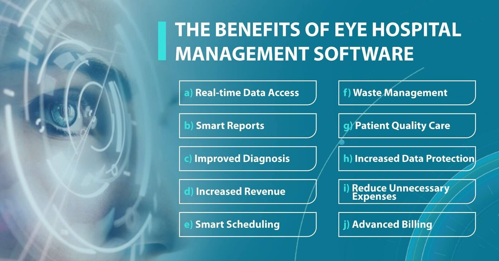
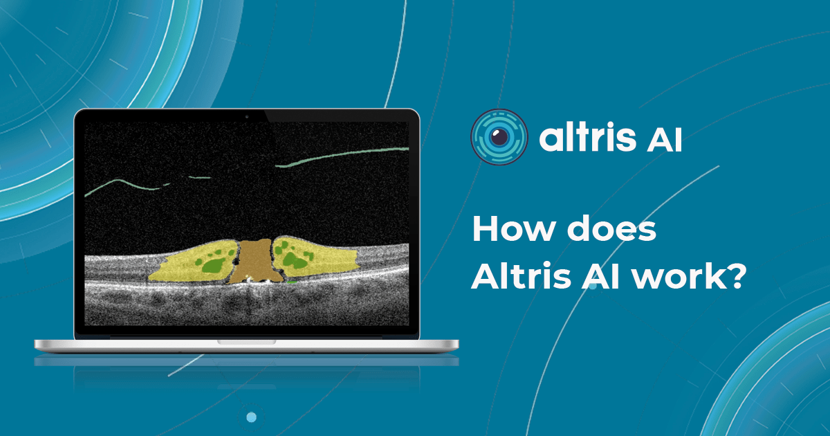


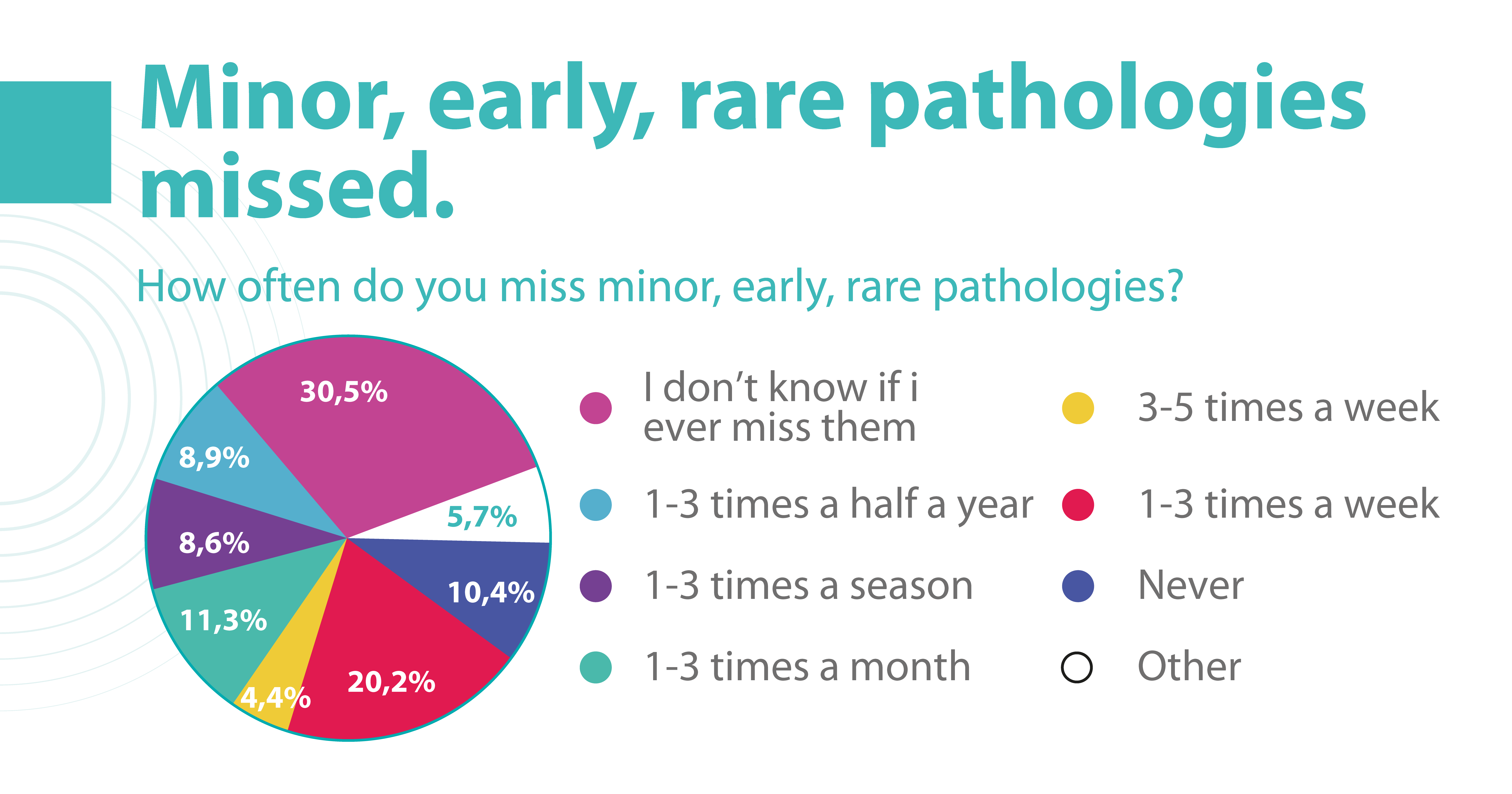

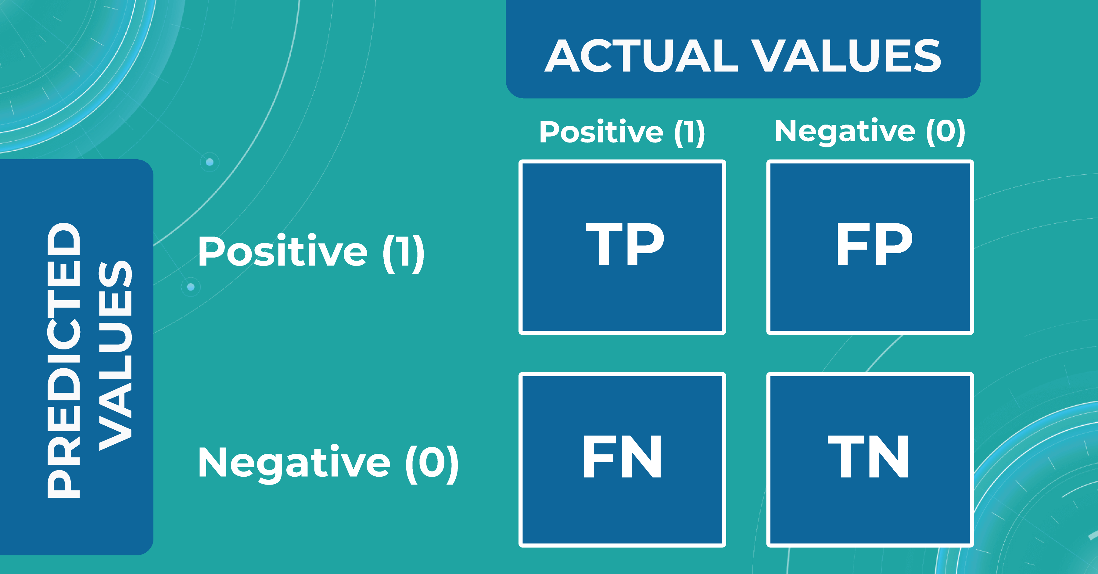


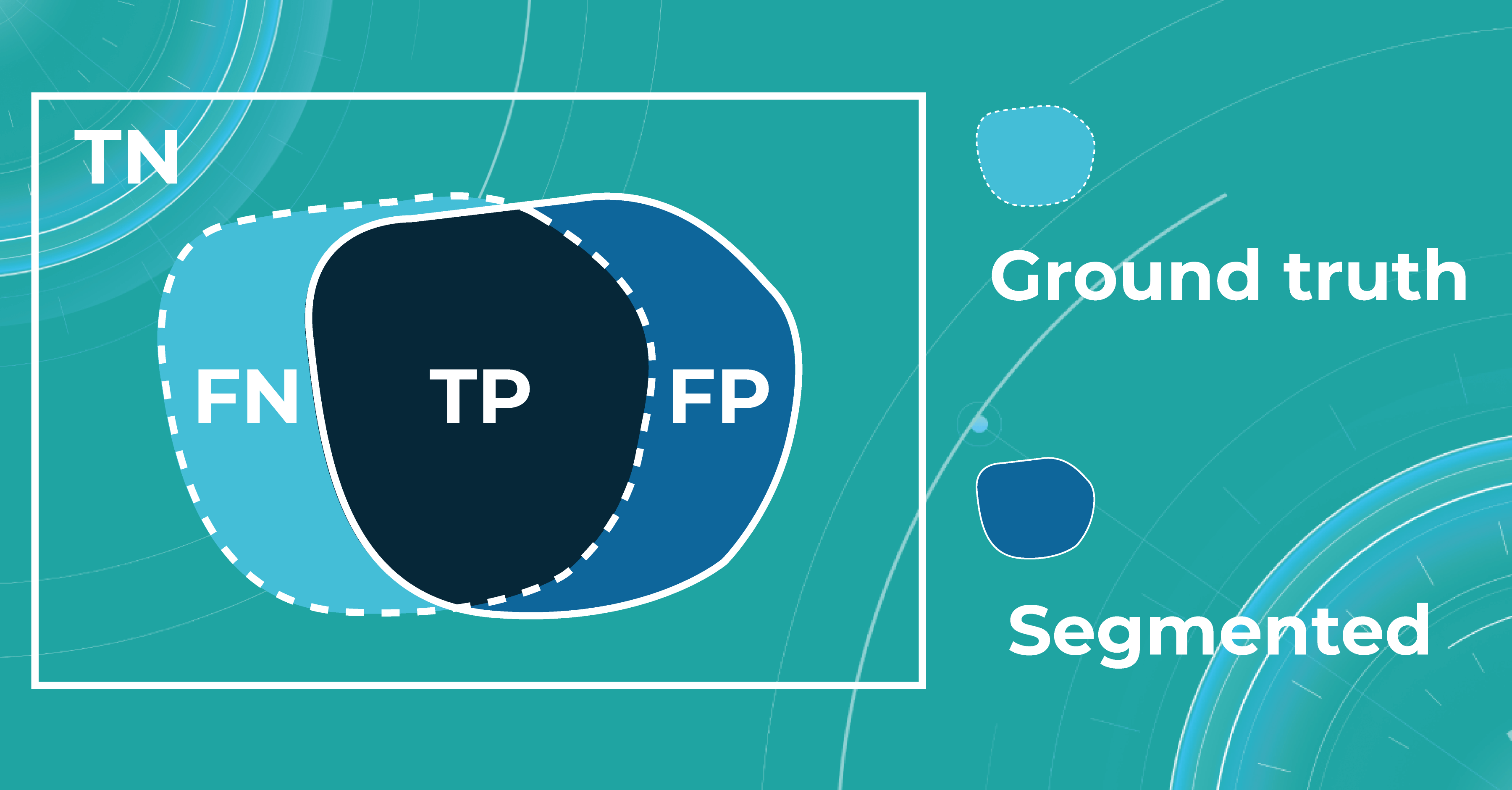
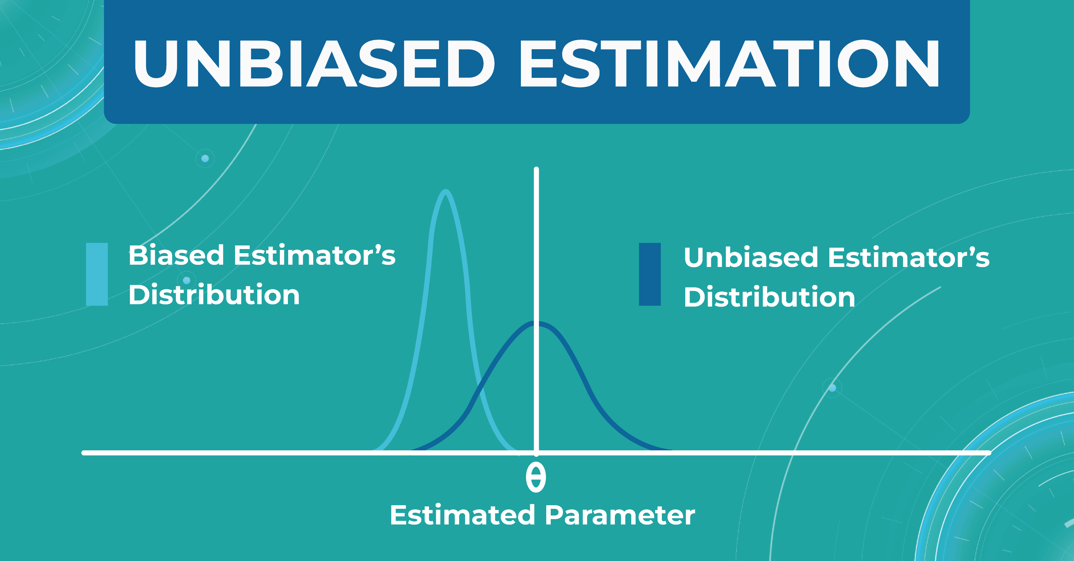
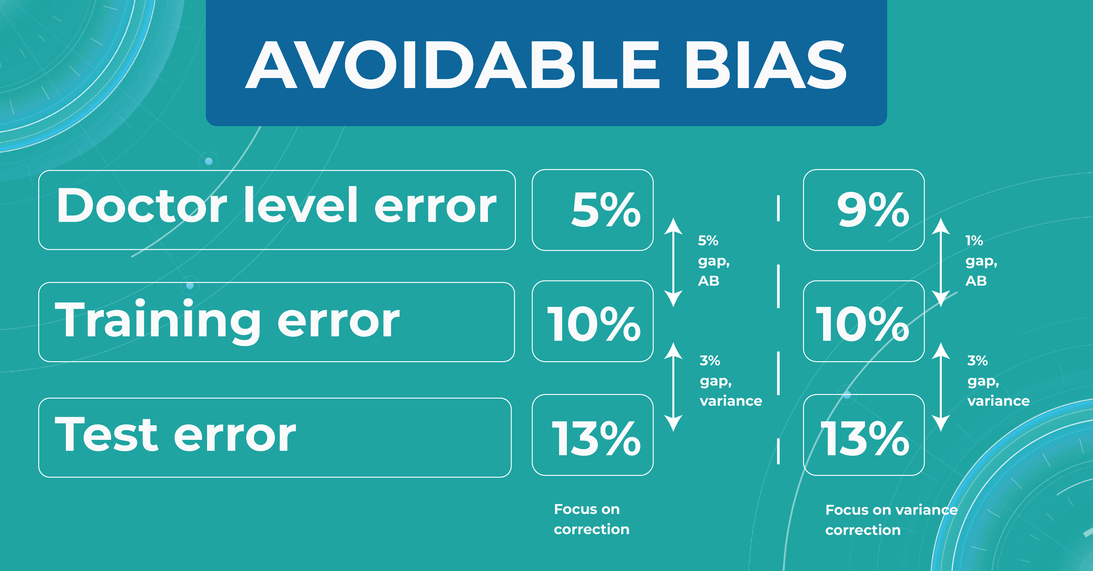
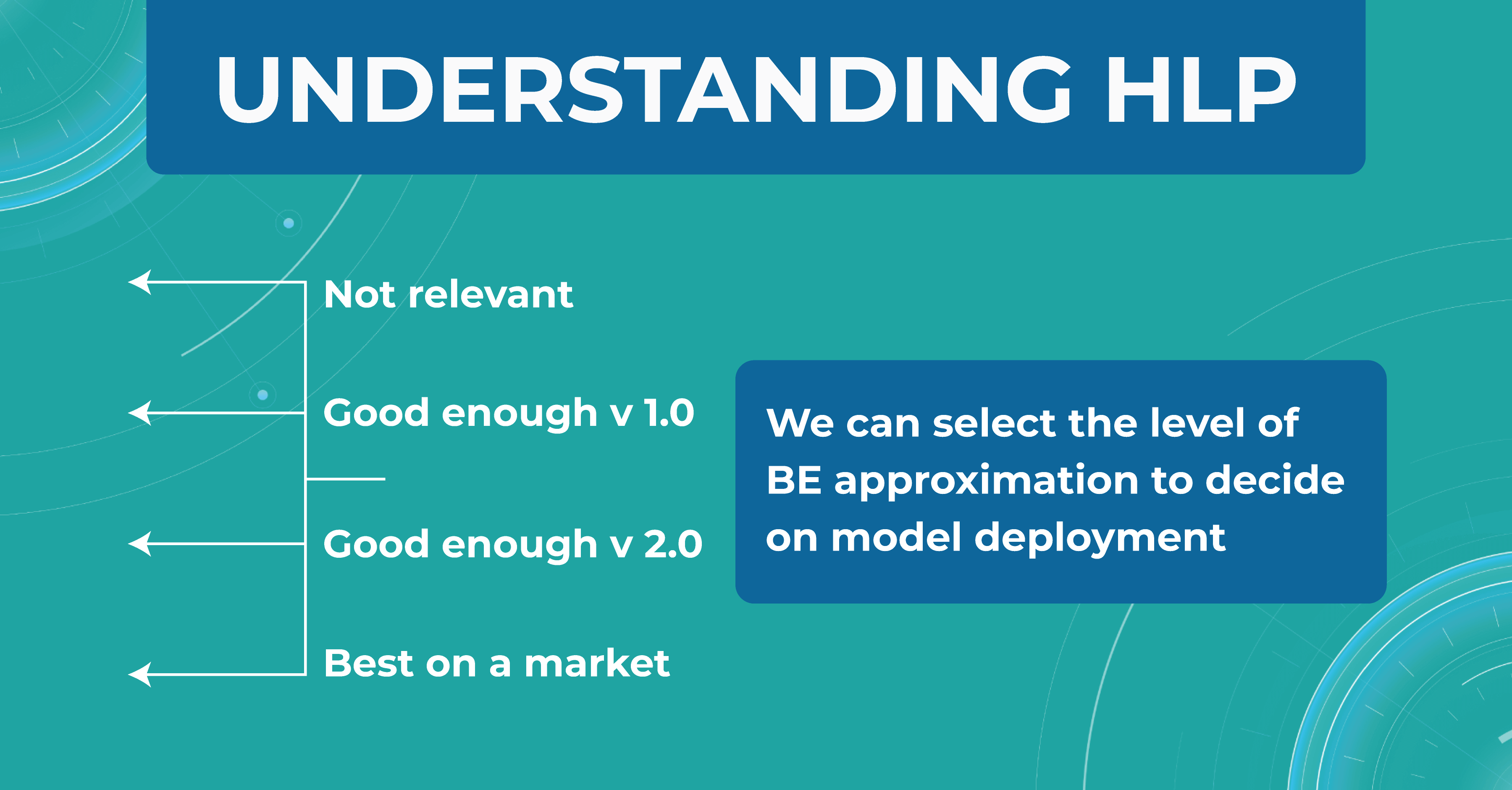
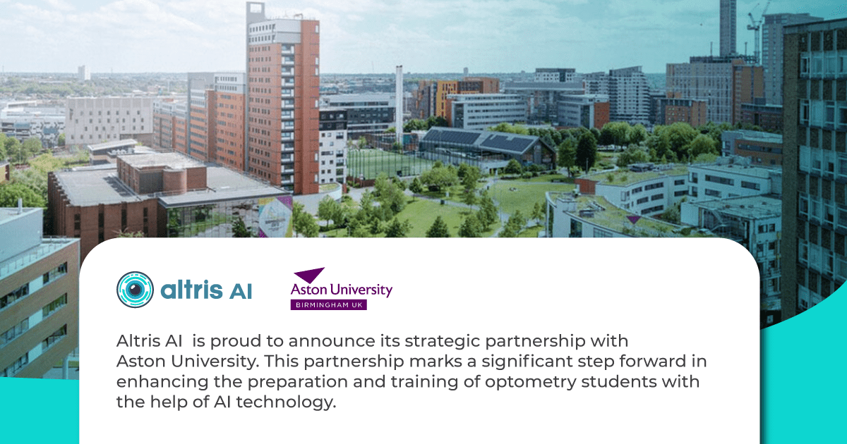
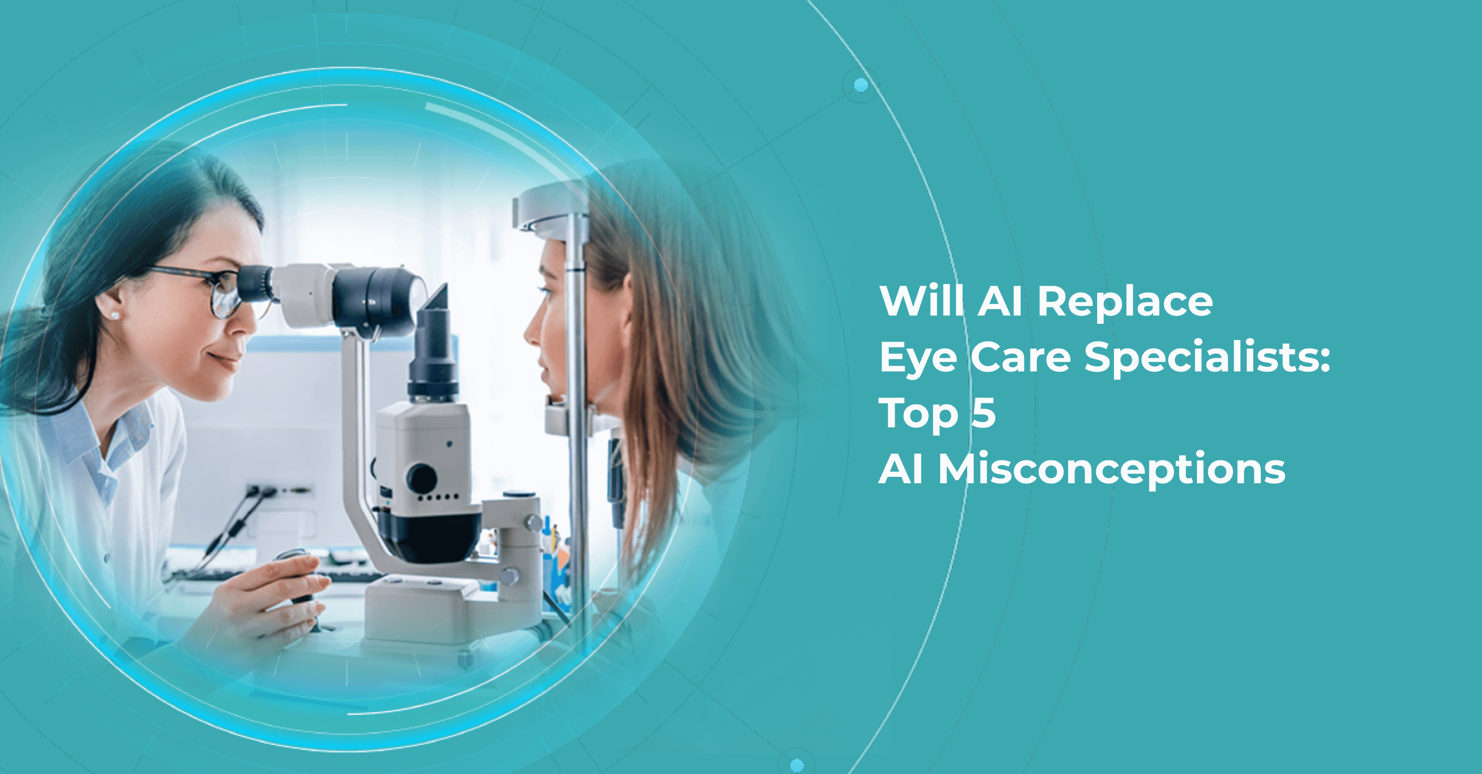


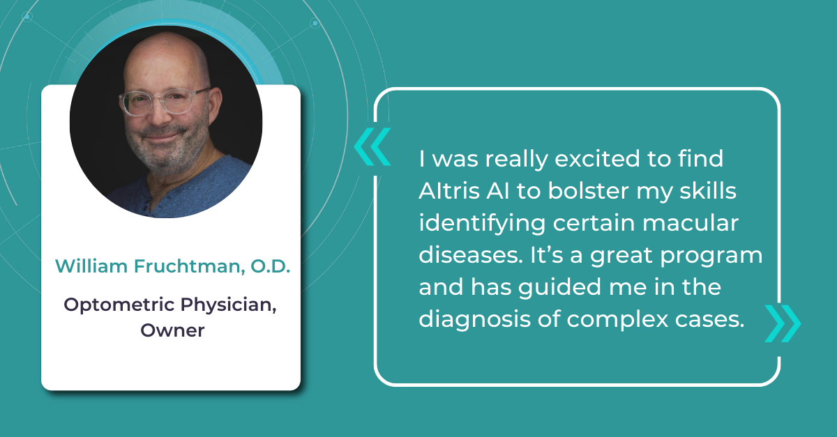

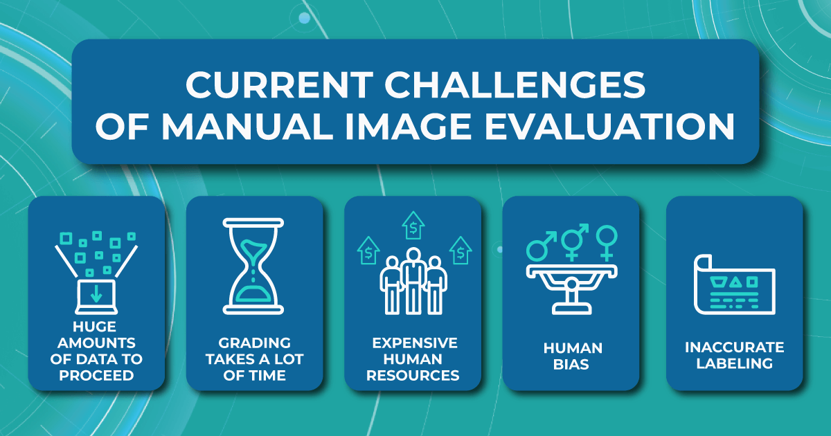
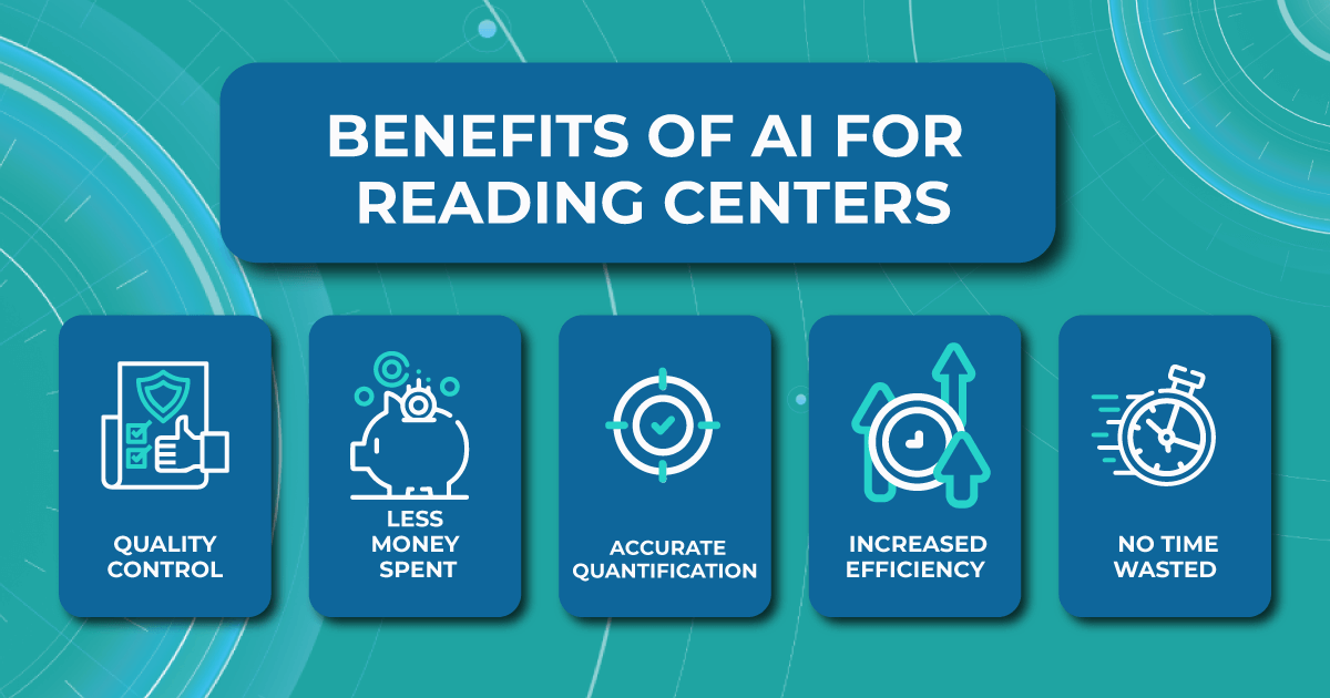
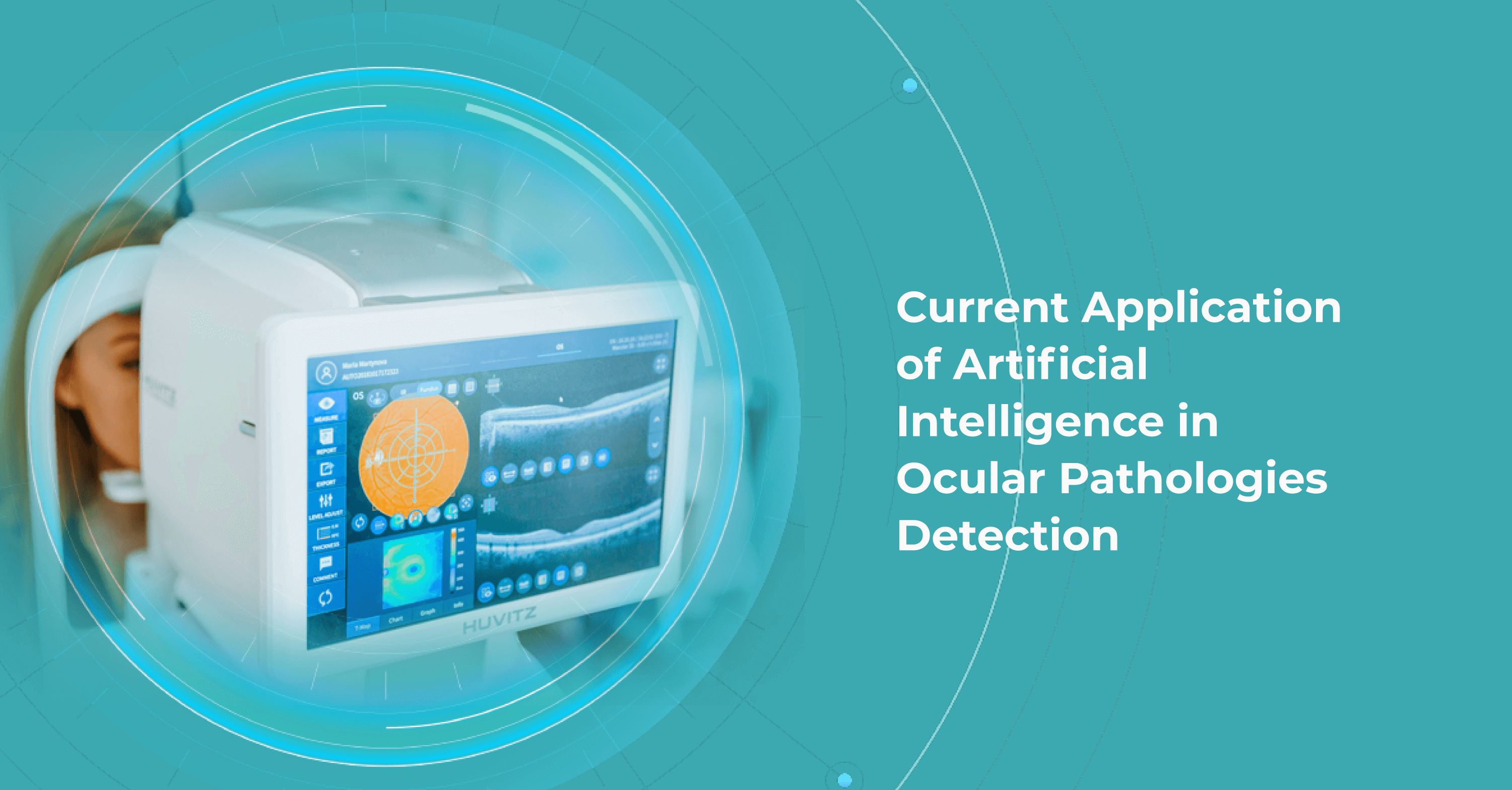
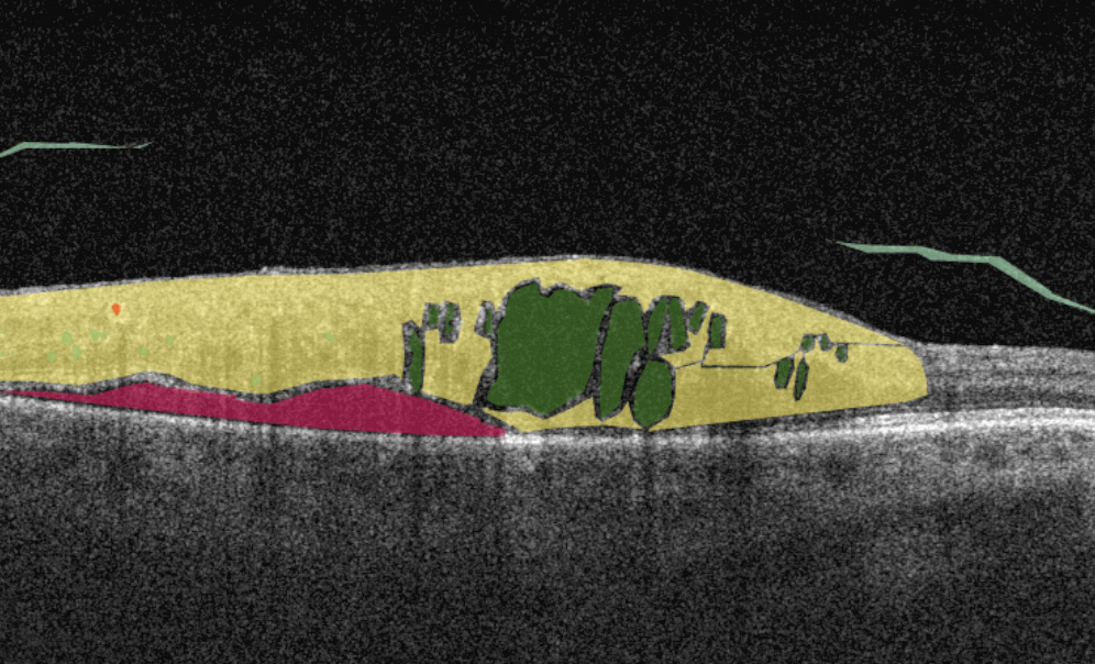
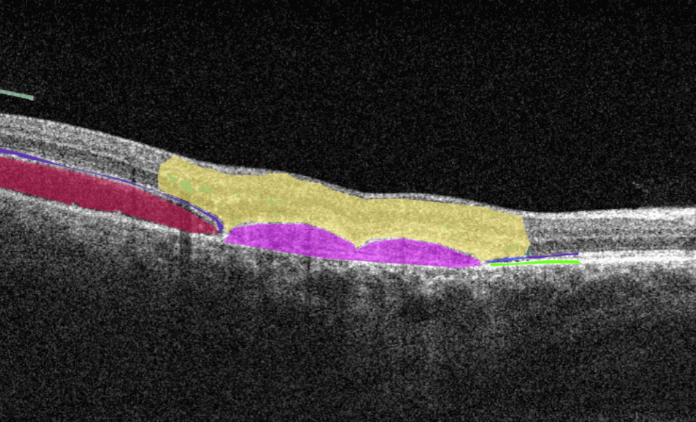
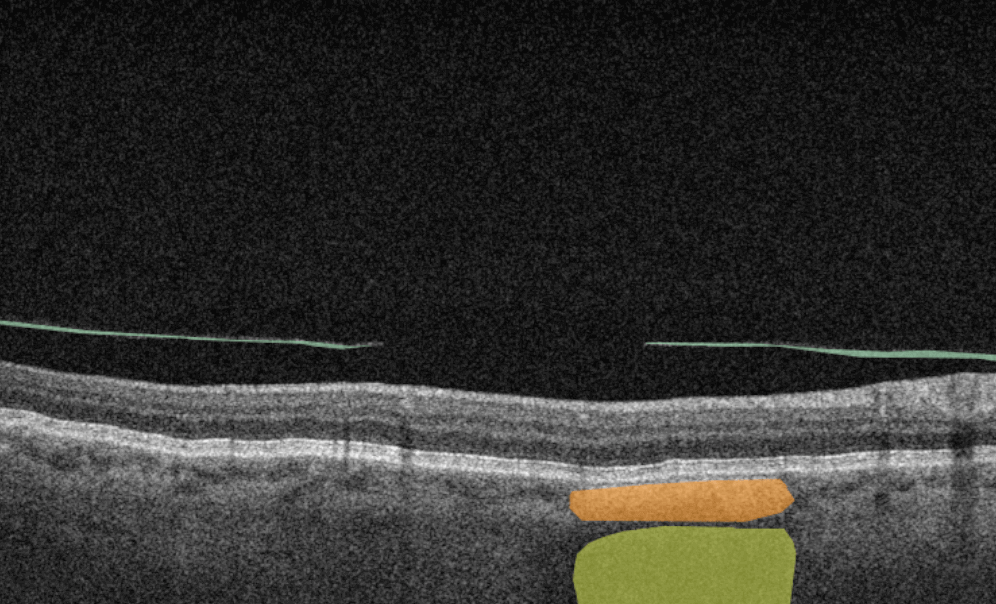
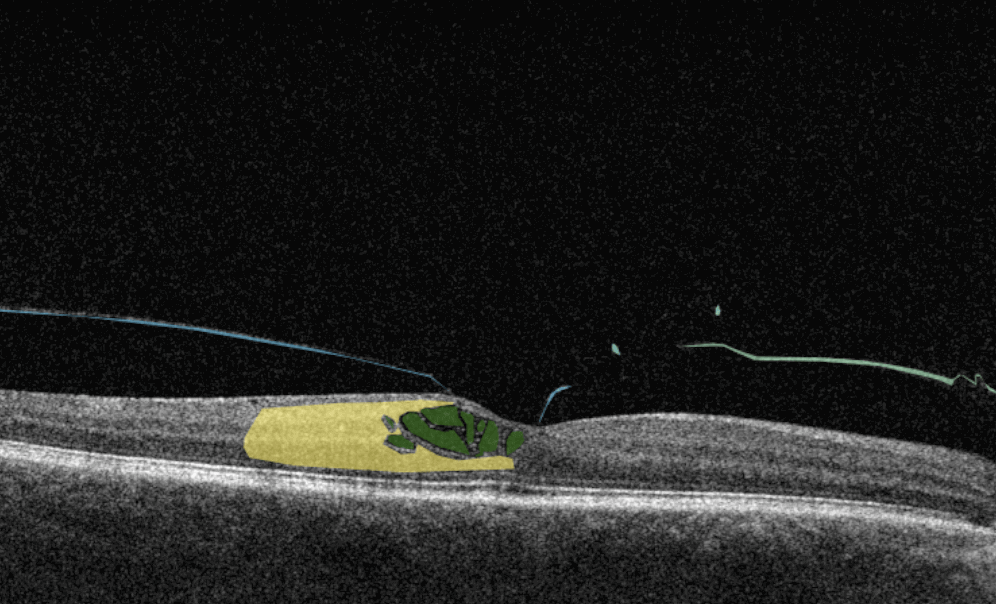
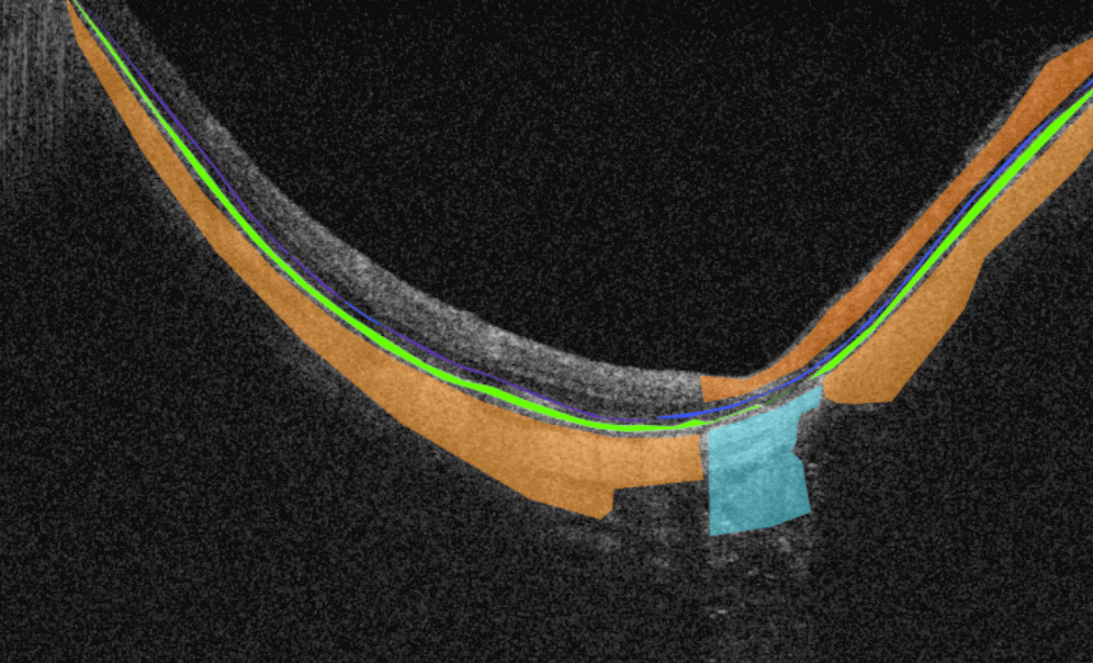
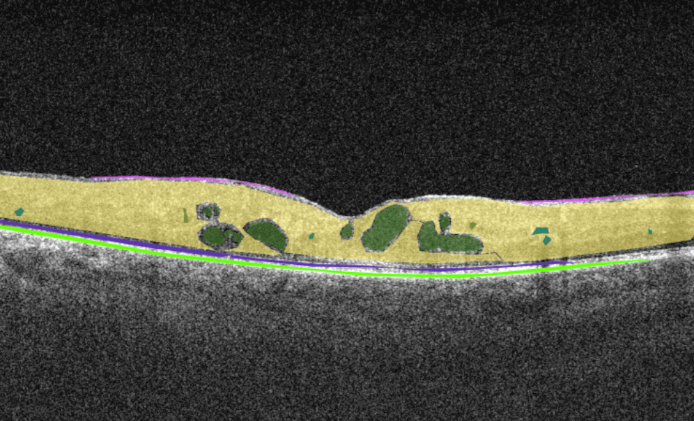

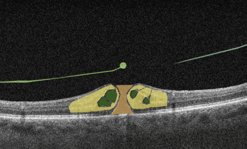
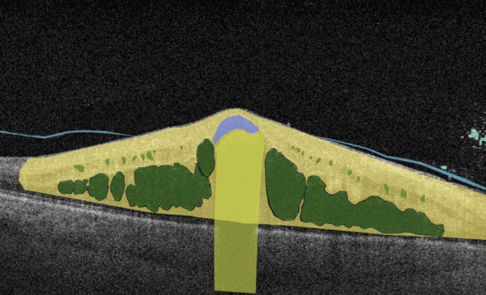
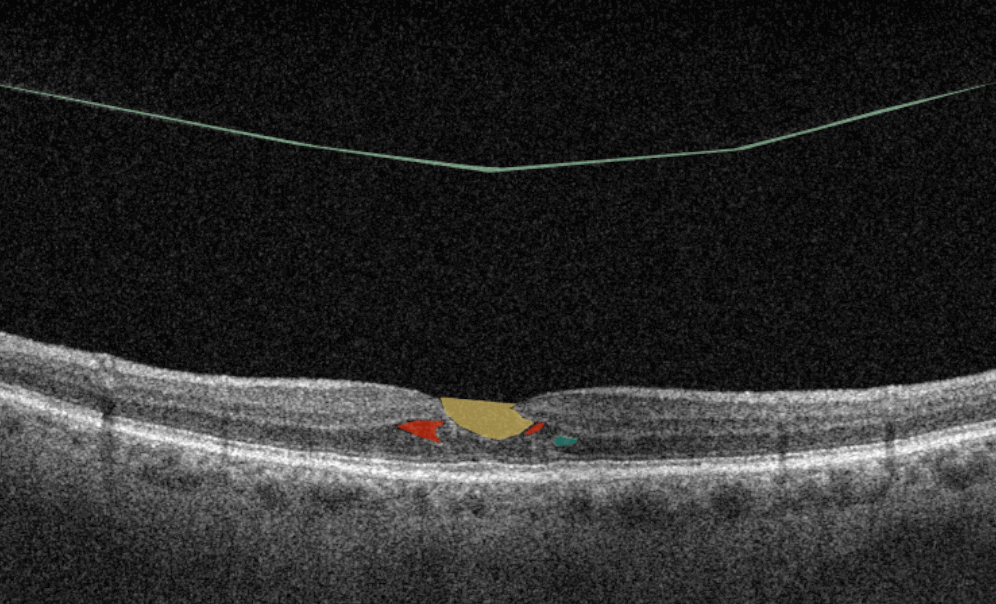
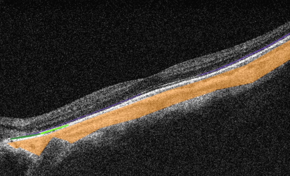
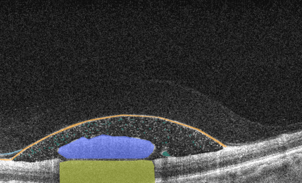
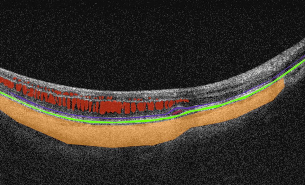
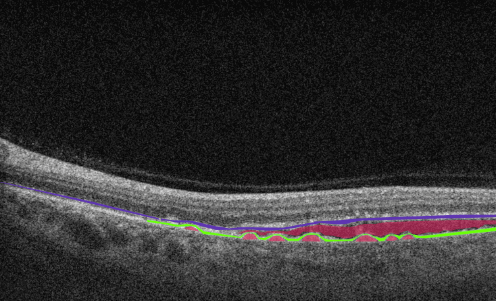