Free Trial

Altris IMS for OCT Ophthalmology Research

Guidance on OCT workflow for ophthalmology research
background
maria

Maria Znamenska, Retina Expert, PhD Ophthalmology, Altris IMS Medical Director

OCT Image Review Workflow: How It Works

My name is Maria Znamenska. I am an ophthalmologist with a PhD in ophthalmology and the Medical Director of Altris IMS. I have been practicing and teaching OCT for more than 16 years. I want to show you how artificial intelligence can be used for OCT analysis with real OCT scans of AMD (drusen), DR, and GA retina pathologies.

OCT is a widely used imaging modality in modern ophthalmic practice. An OCT workflow can assist clinicians by providing structured visualization and quantitative reference information for complex OCT scans, supporting review of retinal features and longitudinal assessment of imaging data. This approach may help streamline OCT review workflows and support consistency in image interpretation.

Researchers' testimonials

Altris IMS for OCT Image Review and Data Research


OCT Research is 1 click away

Free Trial
background

OCT Image Review Workflow for Ophthalmology Research

  • pathology

    When reviewing an OCT scan that requires further evaluation, the system performs an automated analysis of the scan and identifies image features corresponding to 40+ retinal biomarkers in relation to 30+ conditions research. These features are visualized using color overlays to support structured review.

  • measure

    Each biomarker can be automatically quantified using segmentation-based measurements of volume and area, providing standardized quantitative outputs to support image review and analysis.

  • ga
    OCT workflow

    The Measurements module includes functionality for automatically calculating GA-related area measurements based on the spatial overlap of image features associated with hypertransmission and RPE atrophy, with an optional inclusion of neurosensory retina atrophy, as described in research literature.

    For longitudinal review, the Pathology Progression module provides visualization of GA-related area measurements across multiple OCT examinations to support comparative analysis over time.

  • progression
    A green magnifying glass with its lens showing a checkmark inside concentric circles, symbolizing verification or approval. The illustration, suggestive of AI in optometry, is simple with clean lines and a white background.

    The Progression Analysis module enables inclusion of OCT examinations from multiple visits in chronological order and provides visualization of quantitative changes in retinal biomarkers and research-described pathologies.

  • GCC

    Analysis of GCC asymmetry is available to support the evaluation of retinal features associated with glaucoma in research studies.

  • disk area
    OCT workflow

    The Advanced Optic Disc Analysis module measures key optic disc parameters—disc area, cup area, cup volume, cup depth, cup/disc area ratio, and rim absence angle—and generates quantitative outputs intended for research and structured review of OCT data.

  • report
    Examination results can be exported or printed as a customizable report, providing basic information, segmentation data, and visualization of retinal biomarkers and image features studied in research to support organized OCT workflow.

Choose a patient's journey

OCT workflow for analysis of retinal features associated with diabetic retinopathy in research or image review contexts

Diabetic retinopathy (DR) is a retinal microvascular condition associated with diabetes mellitus, which has been extensively studied in research due to its impact on retinal structure and function

  • hard exudates
    A green magnifying glass with its lens showing a checkmark inside concentric circles, symbolizing verification or approval. The illustration, suggestive of AI in optometry, is simple with clean lines and a white background.

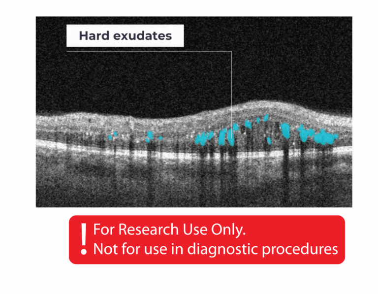
    Hard exudates are deposits composed of lipid and protein-based material, including fibrinogen and albumin, which can accumulate in the retina when the blood–retinal barrier is altered, as described in research studies.

  • diffuse edema
    A green magnifying glass with its lens showing a checkmark inside concentric circles, symbolizing verification or approval. The illustration, suggestive of AI in optometry, is simple with clean lines and a white background.

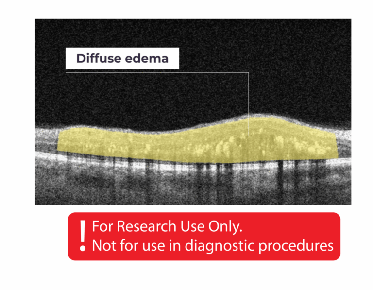
    Diffuse edema is characterized by a thickened region of lower reflectivity in the outer retina, without the presence of cystoid spaces, as described in research literature.

  • foci
    A green magnifying glass with its lens showing a checkmark inside concentric circles, symbolizing verification or approval. The illustration, suggestive of AI in optometry, is simple with clean lines and a white background.

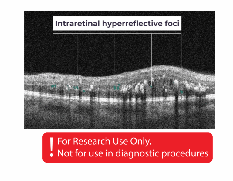
    Intraretinal hyperreflective foci are typically dot-like or round, regularly shaped features observed throughout the retinal layers and choroid, as described in research studies.

  • cystoid fluid
    A green magnifying glass with its lens showing a checkmark inside concentric circles, symbolizing verification or approval. The illustration, suggestive of AI in optometry, is simple with clean lines and a white background.

    Intraretinal cystoid fluid refers to fluid within the retina that appears as cystic cavities, as described in research literature.

OCT workflow for analysis of retinal features associated with dry AMD (drusen) in research or image review contexts

A larger number of prominent drusen can be observed in the retina and has been described in research studies as associated with dry age-related macular degeneration (AMD)

  • Biomarker of dAMD, Soft drusen
    A green magnifying glass with its lens showing a checkmark inside concentric circles, symbolizing verification or approval. The illustration, suggestive of AI in optometry, is simple with clean lines and a white background.

    Soft drusen are generally larger, tend to cluster together, and have edges that are less clearly defined, as described in research studies.

  • OCT Workflow
    A green magnifying glass with its lens showing a checkmark inside concentric circles, symbolizing verification or approval. The illustration, suggestive of AI in optometry, is simple with clean lines and a white background.

    RPE disruption refers to alterations in the structure of the retinal pigment epithelium (RPE) layer, as described in research studies.

  • OCT Workflow
    A green magnifying glass with its lens showing a checkmark inside concentric circles, symbolizing verification or approval. The illustration, suggestive of AI in optometry, is simple with clean lines and a white background.

    Cuticular drusen are small, have steep sides, and contain dense hyalinized material, similar in appearance to small, hard drusen, as described in research studies.

  • Biomarker of dAMD, Hard drusen
    A green magnifying glass with its lens showing a checkmark inside concentric circles, symbolizing verification or approval. The illustration, suggestive of AI in optometry, is simple with clean lines and a white background.

    Hard drusen are small deposits observed in the retina, as described in research studies.

  • AI for OCT detection of Ellipsoid zone disruption
    A green magnifying glass with its lens showing a checkmark inside concentric circles, symbolizing verification or approval. The illustration, suggestive of AI in optometry, is simple with clean lines and a white background.

    The ellipsoid zone is a hyperreflective band in the outer retina, located just posterior to the external limiting membrane, and is commonly evaluated in research studies for its relationship with retinal structure and function.

OCT workflow for analysis of retinal features associated with dry AMD (geographic atrophy) in research or image review contexts

Geographic atrophy (GA) is an advanced form of age-related macular degeneration (AMD), characterized by atrophic lesions in the outer retina that can expand to involve the macula and fovea, as described in research studies.

  • OCT workflow
    A green magnifying glass with its lens showing a checkmark inside concentric circles, symbolizing verification or approval. The illustration, suggestive of AI in optometry, is simple with clean lines and a white background.

    The platform provides tools to analyze retinal features associated with Geographic Atrophy (GA) individually. It also includes functionality to calculate the combined GA area and visualize changes in these features across multiple OCT examinations for research and review purposes.

  • OCT Workflow
    A green magnifying glass with its lens showing a checkmark inside concentric circles, symbolizing verification or approval. The illustration, suggestive of AI in optometry, is simple with clean lines and a white background.

    Hypertransmission can be one of the biomarkers of GA.

  • AI for OCT detection of Hard drusen
    A green magnifying glass with its lens showing a checkmark inside concentric circles, symbolizing verification or approval. The illustration, suggestive of AI in optometry, is simple with clean lines and a white background.

    Hard drusen are small, and indicate lower risk of future vision loss.

  • AI for OCT detection of Soft drusen
    A green magnifying glass with its lens showing a checkmark inside concentric circles, symbolizing verification or approval. The illustration, suggestive of AI in optometry, is simple with clean lines and a white background.

    Soft drusen are larger, cluster together, and have edges that are not as clearly defined.

  • AI for OCT detection of Ellipsoid zone disruption
    A green magnifying glass with its lens showing a checkmark inside concentric circles, symbolizing verification or approval. The illustration, suggestive of AI in optometry, is simple with clean lines and a white background.

    Ellipsoid zone disruption refers to alterations in the hyperreflective outer retinal band located just posterior to the external limiting membrane. These features are commonly evaluated in research studies for their relationship with retinal structure and function.

  • AI for OCT detection of Neurosensory retina atrophy
    A green magnifying glass with its lens showing a checkmark inside concentric circles, symbolizing verification or approval. The illustration, suggestive of AI in optometry, is simple with clean lines and a white background.

    Neurosensory retina atrophy – the atrophy of the inner layer.

  • AI for OCT detection of RPE atrophy
    A green magnifying glass with its lens showing a checkmark inside concentric circles, symbolizing verification or approval. The illustration, suggestive of AI in optometry, is simple with clean lines and a white background.

    RPE Atrophy – the atrophy of Retinal Pigment Epithelium layer.

  • AI for OCT detection of RPE disruption
    A green magnifying glass with its lens showing a checkmark inside concentric circles, symbolizing verification or approval. The illustration, suggestive of AI in optometry, is simple with clean lines and a white background.

    RPE Disruption – the disruption of the Retinal Pigment Epithelium layer.

nAMD(CNV)

  • AI for OCT detection of Ellipsoid zone disruption
    A green magnifying glass with its lens showing a checkmark inside concentric circles, symbolizing verification or approval. The illustration, suggestive of AI in optometry, is simple with clean lines and a white background.

    Ellipsoid Zone Disruption is a hyperreflective outer retinal band just posterior to the external limiting membrane. Its integrity is correlating with visual acuity and other aspects of retinal function.

  • AI for OCT detection of Posterior hyaloid membrane detachment
    A green magnifying glass with its lens showing a checkmark inside concentric circles, symbolizing verification or approval. The illustration, suggestive of AI in optometry, is simple with clean lines and a white background.

    Posterior Hyaloid Membrane Detachment occurs when the retinal layer and vitreous body/posterior hyaloid membrane dissociate, with an intervening fluid collection forming in the subhyaloid space.

  • AI for OCT detection of Subretinal hyperreflective material
    A green magnifying glass with its lens showing a checkmark inside concentric circles, symbolizing verification or approval. The illustration, suggestive of AI in optometry, is simple with clean lines and a white background.

    SHRM is a morphologic component seen as hyperreflective material that is external to the retina and internal to the retinal pigment epithelium (RPE).

  • AI for OCT detection of Diffuse edema
    A green magnifying glass with its lens showing a checkmark inside concentric circles, symbolizing verification or approval. The illustration, suggestive of AI in optometry, is simple with clean lines and a white background.

    Diffuse Edema is a thickened area of lower reflectivity in the outer retina but specifically without cystoid spaces.

  • AI for OCT detection of Fibrovascular RPE detachment
    A green magnifying glass with its lens showing a checkmark inside concentric circles, symbolizing verification or approval. The illustration, suggestive of AI in optometry, is simple with clean lines and a white background.

    Fibrovascular RPE detachment. Delamination of the pigment epithelium of the retina is caused by the presence of newly formed vessels (fibrovascular membrane) under the RPE.

  • AI for OCT detection of Subretinal fluid
    A green magnifying glass with its lens showing a checkmark inside concentric circles, symbolizing verification or approval. The illustration, suggestive of AI in optometry, is simple with clean lines and a white background.

    Subretinal Fluid corresponds to the accumulation of a clear or lipid-rich exudate (serous fluid) in the subretinal space.

  • AI for OCT detection of Double layer sign
    A green magnifying glass with its lens showing a checkmark inside concentric circles, symbolizing verification or approval. The illustration, suggestive of AI in optometry, is simple with clean lines and a white background.

    Double layer sign is a biomarker that is produced due to the shallow irregular pigment epithelial detachment.

  • AI for OCT detection of RPE disruption
    A green magnifying glass with its lens showing a checkmark inside concentric circles, symbolizing verification or approval. The illustration, suggestive of AI in optometry, is simple with clean lines and a white background.

    RPE Disruption is the disruption of the Retinal Pigment Epithelium layer.

maria
Maria Znamenska
MD, PhD Ophthalmology, Medical Director of Altris AI
With Altris IMS, the OCT workflow is designed to support structured review and analysis of retinal images. The system assists in organizing and visualizing OCT scans, highlighting image features associated with biomarkers studied in research. The Progression Analysis module provides quantitative visualization of changes in these features across multiple examinations to support longitudinal review and research-based assessment.
Early changes in glaucoma can be subtle and may be challenging to observe. The Ganglion Cell Complex (GCC) Asymmetry Analysis module provides quantitative visualization of retinal features studied in research. The Glaucoma Early Risk Assessment module supports structured review of these features across OCT scans and provides reference outputs for research and image interpretation.

See how it works in a Demo

Demo Account
background

OCT analysis provides ophthalmologists with tools to enhance the organization, visualization, and review of OCT scans. While OCT imaging is a widely used method for examining the retina, interpreting subtle features can require extensive experience. An OCT workflow supports structured review of retinal images, providing quantitative outputs and visualization of biomarkers to aid research and image interpretation.


  • Altris IMS visualizes over 40 retinal biomarkers and 30 research-described pathologies
  • Altris IMS supports quantitative analysis of retinal features associated with glaucoma in research studies
  • Altris IMS enables longitudinal analysis of retinal biomarkers across multiple OCT scans
  • Altris IMS provides tools to organize and explore retinal imaging data for research purposes via customisable reports
Altris IMS can support ophthalmologists by providing tools for structured review of OCT scans, quantitative analysis of retinal features, and visualization of longitudinal changes in biomarkers studied in research. However, clinical expertise, patient interaction, and nuanced decision-making remain essential aspects of ophthalmology that Altris IMS is not intended to replace.