About Us
Our Team
Compliance
Careers
Product
For Pharma
For Optometry
For Ophthalmology
Cases & Articles
AI for Research
Al for Diabetic Retinopathy
AI for Dry AMD
AI for Wet AMD
AI for Geographic Atrophy
AI for Fluids Research
AI for Glaucoma Research
Free Trial
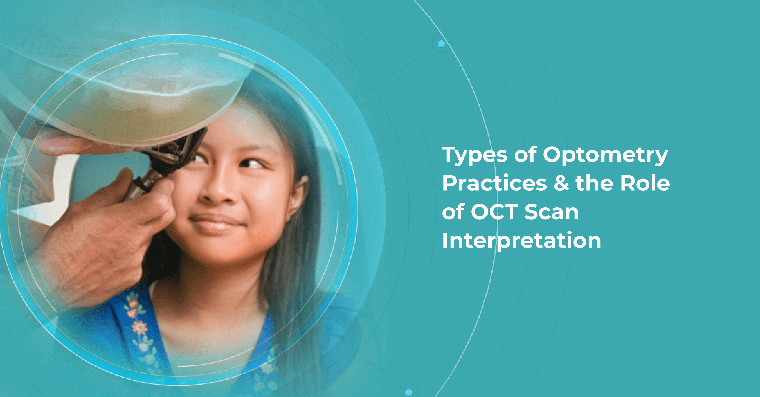
Mark Braddon
VP of Sales at Altris AI
Reading time
7 min. read
We use cookies to ensure that we give you the best experience on our website. If you click "yes" you consent to our use of cookies. Here is our
Privacy policy
Ok
No